
Choose MALDI imaging when the biological conclusion depends on where a lipid is located within tissue. Choose bulk LC-MS lipidomics when the priority is deeper molecular coverage, chromatographic separation, structural confirmation, or quantitative comparison across many samples. Use both when location and molecular confidence are equally important.
This distinction matters because a tissue homogenate averages every compartment it contains. A lipid change confined to a small lesion, anatomical layer, vascular region, or treatment boundary may be diluted or even reversed in bulk data. Conversely, a visually convincing ion image does not by itself establish an isomer-resolved identity or an absolute tissue concentration.
MALDI Imaging and LC-MS Lipidomics at a Glance
- Spatial question: MALDI mass spectrometry imaging (MALDI-MSI) preserves x-y coordinates and maps ion intensity across a tissue section.
- Bulk question: LC-MS analyzes an extracted sample and reports the aggregate lipid composition of the submitted tissue or dissected region.
- Identification: LC separation and MS/MS generally provide stronger support for resolving co-isolated species; an imaging m/z should not automatically be treated as one unique lipid structure.
- Quantification: Routine MALDI images are commonly relative. Quantitative MSI requires internal standards, calibration, matrix-effect assessment, and a validated reporting model.
- Sample design: MALDI-MSI usually favors carefully handled fresh-frozen sections and coordinated histology. Bulk LC-MS accepts a wider range of homogenized tissue designs.
- Best combined design: Use MALDI-MSI to locate molecularly distinct regions and LC-MS/MS on matched, serial, or microdissected material to deepen identification and quantification.
What Do MALDI Imaging and Bulk LC-MS Lipidomics Measure?
MALDI-MSI collects a mass spectrum at each position in a predefined tissue raster. The resulting intensity map associates an m/z signal with its spatial distribution. Pixel spacing determines the acquisition grid, while effective biological resolution also depends on laser footprint, matrix crystal size, analyte diffusion, tissue morphology, and image registration (Moore and Charkoftaki, 2023; Jha et al., 2024).
Bulk LC-MS begins by extracting lipids from a tissue portion. Chromatography separates compounds over time before mass analysis, improving the ability to distinguish lipid classes, isobars, and some isomers while reducing direct competition among coexisting species. The result represents the average composition of everything included in the extraction.
The methods therefore answer different primary questions:
| Study question | MALDI imaging lipidomics | Bulk LC-MS lipidomics |
|---|---|---|
| Where is an ion enriched? | Direct spatial map | Not retained after homogenization |
| Which tissue region differs? | ROI and pixel-level comparison | Requires prior dissection or microdissection |
| What is the overall lipid profile? | Possible but influenced by local ionization and sampling | Directly suited to homogenate-level profiling |
| Can chromatographic retention support identity? | No chromatographic dimension in standard MALDI-MSI | Yes |
| Can many cohort samples be compared quantitatively? | Possible with rigorous normalization and design | Usually the more direct framework |
| Can histology be correlated with chemistry? | Yes, through same-section or serial-section registration | Only indirectly through region selection |
Neither method is inherently more comprehensive. Spatial completeness and molecular completeness are different dimensions. A project can map hundreds of ions yet have limited structural certainty, or identify many lipids precisely while losing their tissue localization.
The site's introductory MALDI imaging lipidomics guide explains the imaging principle. The decision in a new project should start one level earlier: whether tissue position is part of the hypothesis or merely useful context.
When Does Spatial Context Change the Lipidomics Conclusion?
Spatial context matters when averaging tissue compartments would obscure the biological contrast. It is most valuable when the tissue contains known or suspected molecular neighborhoods that cannot be separated reliably before analysis.
Anatomical layers and small regions of interest
Brain nuclei, renal cortex and medulla, retinal layers, epidermal and dermal compartments, tumor core and margin, necrotic regions, and plant vascular structures can contain distinct lipid programs. A bulk measurement can show that a lipid changed, but it cannot reveal whether the signal originated from one layer or shifted between neighboring regions.
MALDI-MSI is appropriate when the planned comparison is region-to-region within a section or when an unanticipated molecular region may emerge from unsupervised segmentation. Histology, optical imaging, or molecular markers should be incorporated so that ion clusters are interpreted against actual anatomy rather than image appearance alone.
Local gradients and treatment distribution
Spatial imaging can reveal gradients around a delivery site, lesion boundary, implant, vascular feature, or exposure interface. These patterns may support hypotheses about transport, metabolism, or tissue response that cannot be reconstructed from one homogenized value.
The study still needs biological replication. Thousands of pixels in one section are not thousands of independent biological samples. Pixel-level statistics must respect the hierarchy of pixels within regions, sections, and experimental subjects.
Molecular heterogeneity inside apparently similar tissue
Histologically similar regions may contain distinct lipid signatures, while visibly different regions may share molecular features. MALDI-MSI can identify spatial clusters without predefined antibodies and can be registered to histology or immunofluorescence. Histology-compatible workflows have demonstrated that lipid imaging and subsequent staining can be coordinated, but the order and compatibility of preparation steps must be validated (Kaya et al., 2017).
For research questions centered on regional tissue biology, the MALDI-Imaging Lipidomics service is the direct analytical route. If the hypothesis only requires a tissue-average group comparison, spatial acquisition may add data volume without changing the scientific decision.
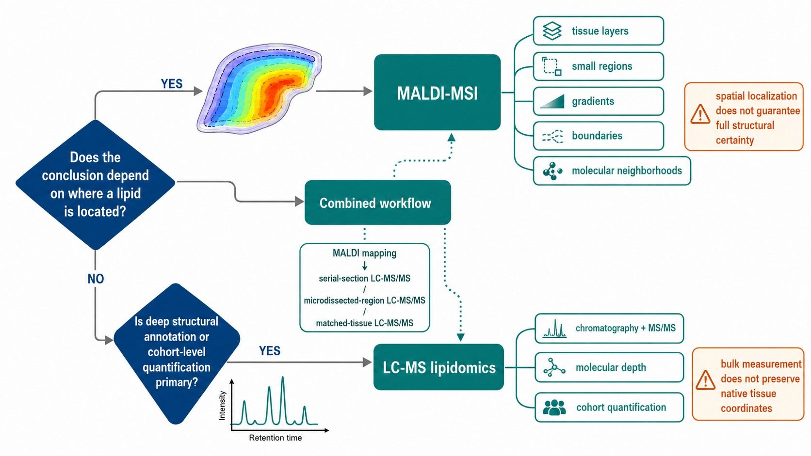 MALDI imaging is selected when spatial context changes interpretation; LC-MS is selected for molecular depth and cohort quantification.
MALDI Imaging vs LC-MS for Lipid Coverage and Identification
The same nominal mass can represent multiple lipid structures, adducts, isotopes, or fragments. This is a central limitation in spatial lipidomics because an attractive ion map can encourage overinterpretation of an m/z assignment.
Why LC-MS usually supports deeper structural annotation
LC-MS can combine accurate mass, retention behavior, isotope pattern, adduct logic, and tandem mass spectra. Targeted methods can add authentic standards and analyte-specific transitions. Specialized separations or fragmentation can further address fatty-acyl composition, sn-position, or double-bond location, although the achievable structural level remains method dependent.
Chromatography also spreads the lipid mixture over time. This reduces spectral congestion and allows selected precursor ions to be interrogated with MS/MS. Bulk LC-MS is therefore usually better suited when the primary deliverable is a large annotated lipid table or a validated panel of named molecular species.
Why MALDI-MSI assignments need orthogonal evidence
Standard MALDI-MSI often starts with accurate m/z and spatial pattern. Tissue salts can create different adducts, abundant lipids can suppress other species, and isobars or isomers can overlap. Database matching alone supports a putative annotation, not a unique molecular identity.
Ion mobility and on-tissue fragmentation can improve confidence. Djambazova et al. demonstrated that MALDI-TIMS increased peak capacity by more than 250% in their system and separated several classes of lipid isomers with different tissue distributions (Djambazova et al., 2020). Even so, the required structural claim should be specified before data acquisition because not every ion will receive equivalent confirmation.
Match the identification level to the biological claim
If the claim is that an ion at a defined m/z is enriched in one region, imaging evidence may be sufficient when reported conservatively. If the claim depends on a particular lipid class or sum composition, accurate mass and MS/MS should support that level. If it depends on acyl chains, double-bond position, stereochemistry, or an absolute concentration, additional separation, standards, or orthogonal analysis may be required.
If structural specificity is central, choose LC-MS or a combined workflow. If localization of a well-characterized ion is central, choose MALDI-MSI with the appropriate confirmation strategy.
MALDI Imaging vs LC-MS for Quantification
Ion intensity is not automatically concentration. In MALDI-MSI, local tissue composition, salt content, matrix deposition, extraction into the matrix layer, laser sampling, and ion suppression can all change signal response across a section.
Relative imaging is different from quantitative MSI
Many imaging studies report normalized relative intensity or regional fold change. This can be scientifically useful when technical variation is controlled and comparisons are made within a qualified design. It should not be described as absolute concentration without calibration and validation.
Quantitative MSI can use isotopically labeled or class-matched standards, tissue-mimetic calibration, standard deposition, and pixel-wise normalization. Vandenbosch et al. applied a multiclass internal-standard mixture to quantify 13 lipid classes and subclasses representing almost 200 sum-composition species in brain tissue, illustrating both the potential and the method development required (Vandenbosch et al., 2023).
Bulk LC-MS provides a more established concentration framework
For defined analytes, LC-MS can use matrix-matched calibration, stable-isotope standards, recovery evaluation, dilution checks, and analyte-specific acceptance criteria. It is usually the stronger choice when the primary endpoint is concentration per tissue mass, protein amount, or another predefined denominator across a cohort.
Bulk analysis is not free from matrix effects, extraction bias, or standard limitations. Its advantage is that these effects can be evaluated within a mature quantitative workflow rather than inferred from raw peak area.
The targeted lipidomics service is appropriate when named lipids and quantitative comparability are primary. If the study requires regional abundance rather than a whole-tissue concentration, quantitative MALDI-MSI or region-guided LC-MS should be considered instead.
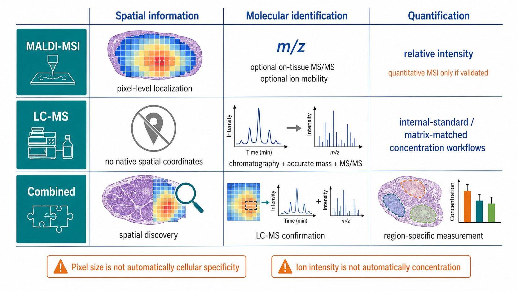 MALDI-MSI and LC-MS produce different forms of spatial, structural, and quantitative evidence.
Tissue Sampling Requirements for MALDI Imaging and LC-MS
Method selection must happen before tissue collection. A sample preserved for routine bulk extraction may no longer support reliable imaging, and a single thin section may not provide enough material for deep LC-MS confirmation.
MALDI imaging sample planning
Fresh-frozen tissue is commonly preferred for endogenous lipid imaging because fixation and embedding materials can extract lipids, introduce background, or alter ionization. Tissue orientation, freezing method, section integrity, section thickness, mounting substrate, and storage history should be standardized.
The region of interest must fit within the section and remain interpretable after matrix application and laser acquisition. Serial sections should be reserved for histology, orthogonal staining, replicate imaging, or LC-MS confirmation. Adjacent sections are similar but not identical, especially when the feature of interest is small.
Matrix selection and application influence sensitivity, ion polarity, spatial resolution, and analyte diffusion. These are analytical design variables, not cosmetic preparation steps. Jha et al. emphasized that tissue handling, matrix application, crystal formation, and lateral diffusion can all affect reproducibility and spatial fidelity (Jha et al., 2024).
Bulk LC-MS tissue planning
Bulk LC-MS requires a clearly defined tissue unit. Whole organ, punch, macrodissected region, laser-capture microdissection, and residual tissue adjacent to an imaging section answer different questions. Record wet mass or another validated input measure, anatomical origin, dissection boundaries, ischemia or handling interval, preservation, and freeze-thaw history.
Tissue homogenization improves representativeness within the selected region but permanently removes spatial information. For heterogeneous tissue, several carefully defined subregions may be more informative than one whole-tissue extract.
The tissue untargeted lipidomics service supports broad extracted-tissue profiling. If a project may later need imaging, retain oriented, intact frozen material and document sectioning plans before allocating all tissue to homogenization.
How to Combine MALDI Imaging and LC-MS Lipidomics
The strongest design is often not a binary choice. MALDI-MSI can discover where molecular differences occur, while LC-MS/MS can strengthen identification and quantify selected regions or matched tissue.
Serial-section design
One section is used for MALDI-MSI, an adjacent section for histology, and another for extraction or region-specific sampling. This design preserves each analytical workflow but assumes that adjacent sections represent the same feature. It is suitable when regions are larger than the distance between sections and tissue architecture is consistent.
MALDI-guided region extraction
Imaging identifies molecularly distinct regions that can be macrodissected or laser-microdissected from a matched section for LC-MS. This converts an unexpected image pattern into a targeted molecular question. The extracted region must provide enough material, and the registration method should be documented.
Same-section sequential analysis
Specialized workflows can combine imaging and extraction from the same tissue section. Hendriks et al. showed the feasibility of integrating quantitative MSI with LC-MS/MS lipidomics and proteomics from one tissue area, while also observing that prior MSI affected subsequent measurements and reduced agreement for some lipid quantifications (Hendriks et al., 2024).
Same-section analysis conserves scarce material but should not be assumed neutral. The sequence of matrix deposition, laser ablation, extraction, and orthogonal analysis must be qualified for the intended endpoint.
Discovery-to-validation design
A subset of representative sections can undergo imaging to define regions and candidate lipids. The larger cohort can then be measured by targeted or untargeted LC-MS using standardized dissections. This approach balances spatial discovery with cohort-scale comparability.
If the study needs exact localization in every sample, image the cohort. If it needs spatial discovery followed by robust group-level measurement, image a representative subset and validate the resulting targets or regions by LC-MS.
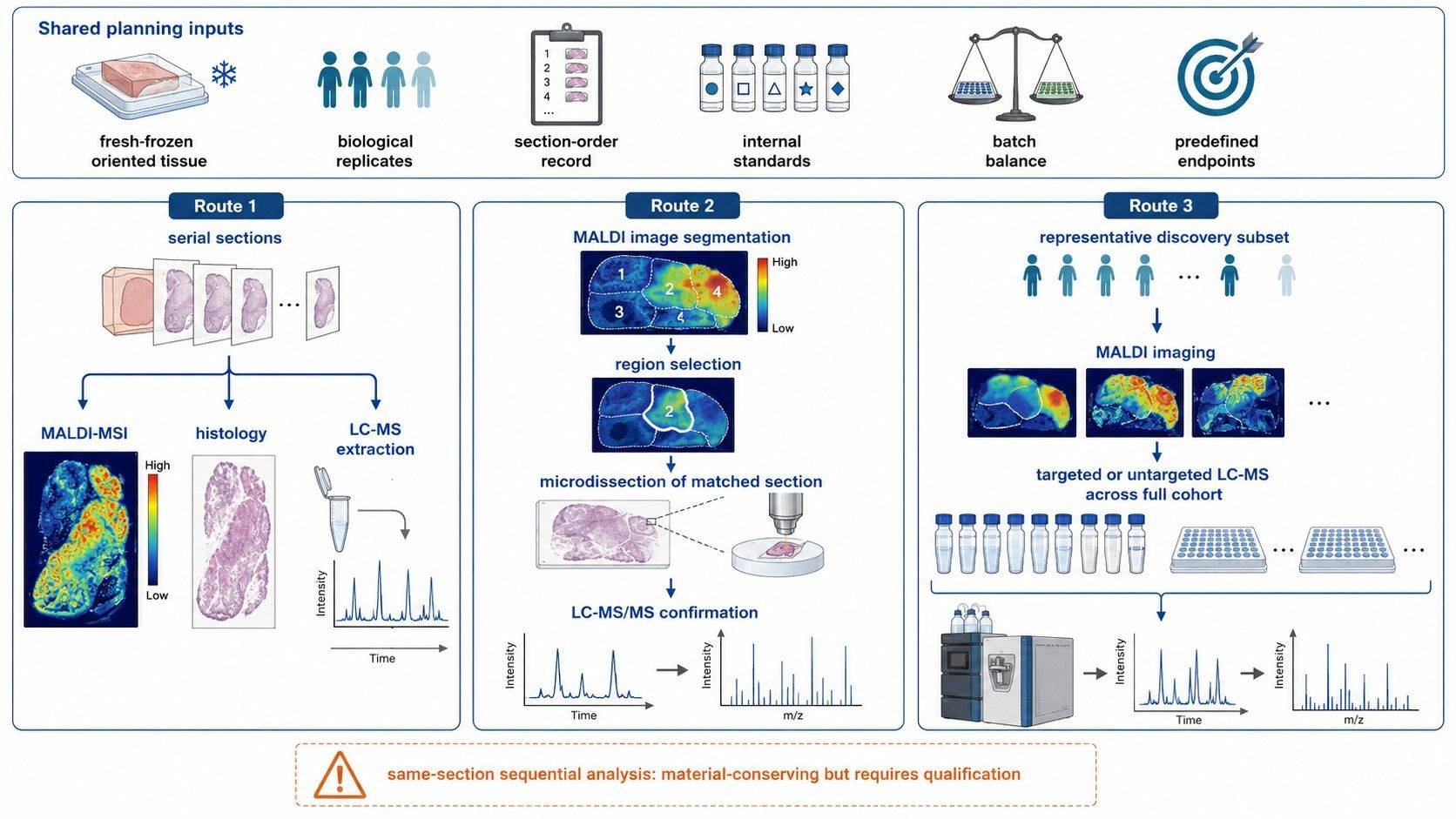 Integrated MALDI imaging and LC-MS designs connect tissue localization with lipid identification and cohort-level measurement.
QC and Replication for Spatial and Bulk Lipidomics
Imaging and bulk workflows need different technical controls, but both must preserve biological replication and separate analytical variance from group effects.
| QC element | MALDI imaging emphasis | Bulk LC-MS emphasis |
|---|---|---|
| Biological replication | Independent tissues, not pixels | Independent tissue extracts |
| Allocation | Balance groups across slides, runs, and preparation batches | Randomize extraction and injection order |
| Blank | Matrix-coated area, substrate, and preparation blank | Solvent, extraction, and carryover blanks |
| Reference material | Serial control section or tissue microarray where feasible | Pooled extract QC and bridge samples |
| Internal standards | Uniform deposition and adduct-compatible response | Early addition, class coverage, recovery, and drift monitoring |
| Image validation | Histology registration, ROI rules, pixel filtering | Peak integration, retention, MS/MS, and annotation criteria |
| Batch assessment | Slide, matrix-coating day, sectioning batch, acquisition order | Extraction plate, column, source, and injection order |
Imaging studies should predefine whether statistics operate on pixels, ROIs, sections, or tissue donors. The independent experimental unit is usually the biological specimen. Treating correlated pixels as independent replicates can produce misleading significance.
For both platforms, group labels should be balanced across preparation and acquisition batches. A reference section or tissue microarray may reveal slide-to-slide drift in imaging; pooled extracts and system suitability samples serve a related role in LC-MS.
Common Errors When Choosing MALDI Imaging or LC-MS
| Design error | Why it fails | Better decision |
|---|---|---|
| Choosing MALDI only because an image is persuasive | The study may not require location, while identification and quantification become secondary | Define the spatial hypothesis and required molecular confidence first |
| Using whole-tissue LC-MS for a small focal feature | The region-specific signal is diluted by surrounding tissue | Image the section or dissect the region before extraction |
| Treating one m/z as one lipid structure | Isobars, isomers, adducts, and fragments may overlap | Report the supported annotation level and add MS/MS, mobility, or LC-MS confirmation |
| Interpreting pixels as biological replicates | Spatial observations within one specimen are correlated | Replicate tissues and use hierarchical statistics |
| Comparing raw MALDI intensity as concentration | Local matrix and ionization effects are uncontrolled | Use validated normalization or quantitative MSI |
| Preparing all tissue for one platform | The complementary question becomes impossible to answer | Reserve oriented sections and matched tissue during collection |
| Ignoring section order and registration | Small regions may not align across serial sections | Record section sequence and define registration landmarks |
Planning a MALDI Imaging or LC-MS Lipidomics Project
A useful project request should describe the tissue, species or model, preservation and embedding history, number of biological replicates, section dimensions, known anatomical regions, primary lipid classes, expected effect, and whether the endpoint is localization, relative profiling, structural identification, or absolute quantification.
For imaging, also provide the desired region or feature size, available histology, section orientation, whether serial sections exist, and whether the project requires same-section staining or multi-omics registration. For LC-MS, provide tissue amount, dissection strategy, preferred normalization denominator, and whether the study is targeted or untargeted.
Creative Proteomics can align MALDI-MSI, tissue extraction, targeted or untargeted LC-MS, internal standards, histology registration, ROI analysis, and orthogonal confirmation in one study plan. The most informative consultation occurs before all tissue is fixed, embedded, sectioned, or homogenized.
If location determines the interpretation, select MALDI imaging. If molecular depth and cohort-level quantification determine the interpretation, select LC-MS. If the project needs both, reserve matched material and plan the integration before sample collection.
FAQs
Can MALDI imaging replace LC-MS lipidomics?
Not for every objective. MALDI-MSI preserves lipid location, while LC-MS generally provides stronger chromatographic separation, annotation depth, and quantitative workflows. MALDI can replace bulk analysis only when spatial mapping is the primary endpoint and the required identification and quantification can be supported within the imaging method.
Does higher spatial resolution always improve a MALDI lipidomics study?
No. Smaller pixels can increase acquisition time, data volume, and the risk of insufficient ion signal per pixel. Choose a resolution that can distinguish the biological feature of interest while preserving sensitivity, throughput, and reproducibility.
Can MALDI imaging provide absolute lipid concentrations?
Yes, but only through a quantitative MSI method with suitable standards, calibration, normalization, matrix-effect evaluation, and validation. A normalized ion-intensity map alone is a relative distribution, not an absolute concentration map.
How should MALDI ion identities be confirmed?
Use evidence appropriate to the structural claim: accurate mass, isotope and adduct logic, on-tissue MS/MS, ion mobility, authentic standards, and LC-MS/MS of matched tissue or regions. Report annotations at the level the evidence actually supports.
Should the same tissue section be used for imaging and LC-MS?
It can be, but prior matrix deposition and laser sampling may affect the later extraction. Same-section workflows require qualification. Serial sections or matched region extracts are often simpler when sufficient tissue is available.
How many tissue sections are needed?
The number depends on tissue heterogeneity, section quality, polarity modes, histology, technical validation, and biological replication. Plan enough sections for the primary imaging run, adjacent histology, failed-section reserve, and any LC-MS or staining confirmation without treating serial sections as independent biological replicates.
References:
- Djambazova, K. V., Klein, D. R., Migas, L. G., et al. (2020). Resolving the Complexity of Spatial Lipidomics Using MALDI TIMS Imaging Mass Spectrometry. Analytical Chemistry, 92(19), 13290-13297. https://doi.org/10.1021/acs.analchem.0c02520
- Hendriks, T. F. E., Krestensen, K. K., Mohren, R., et al. (2024). MALDI-MSI-LC-MS/MS Workflow for Single-Section Single Step Combined Proteomics and Quantitative Lipidomics. Analytical Chemistry, 96(10), 4266-4274. https://doi.org/10.1021/acs.analchem.3c05850
- Jha, D., Blennow, K., Zetterberg, H., Savas, J. N., & Hanrieder, J. (2024). Spatial Neurolipidomics-MALDI Mass Spectrometry Imaging of Lipids in Brain Pathologies. Journal of Mass Spectrometry, 59(3), e5008. https://doi.org/10.1002/jms.5008
- Kaya, I., Michno, W., Brinet, D., et al. (2017). Histology-Compatible MALDI Mass Spectrometry Based Imaging of Neuronal Lipids for Subsequent Immunofluorescent Staining. Analytical Chemistry, 89(8), 4685-4694. https://doi.org/10.1021/acs.analchem.7b00313
- Moore, J. L., & Charkoftaki, G. (2023). A Guide to MALDI Imaging Mass Spectrometry for Tissues. Journal of Proteome Research, 22(11), 3401-3417. https://doi.org/10.1021/acs.jproteome.3c00167
- Vandenbosch, M., Mutuku, S. M., Mantas, M. J. Q., et al. (2023). Toward Omics-Scale Quantitative Mass Spectrometry Imaging of Lipids in Brain Tissue Using a Multiclass Internal Standard Mixture. Analytical Chemistry, 95(51), 18719-18730. https://doi.org/10.1021/acs.analchem.3c02724
Author: CAIMEI LI, Senior Scientist