Host-Pathogen Interaction Lipidomics

Host-Pathogen Interaction Lipidomics Services

Creative Proteomics provides high-resolution, targeted, and untargeted lipidomics services to decode the complex biochemical arms race between pathogens and their hosts. We empower infection biologists, immunologists, and drug developers to transition from simple phenotypic infection assays to deep mechanistic mapping, delivering reliable quantification of pathogen-induced lipid remodeling, host membrane hijacking, and inflammatory lipid mediator bursts.

Key capabilities

  • Viral Envelope & Membrane Fusion Analysis: Track the exact host lipids (such as specific sphingolipids or cholesterol) hijacked by viruses for viral entry, replication, and envelope budding.
  • Macrophage & Intracellular Pathogen Profiling: Evaluate how intracellular bacteria manipulate host macrophage lipid droplets to secure energy and evade immune clearance.
  • Inflammatory Lipid Mediator Quantification: Measure trace-level pro-inflammatory and pro-resolving eicosanoids (prostaglandins, leukotrienes) during systemic infection or sepsis models.
Request Analysis

Submit Your Request Now

×

Our services have earned the trust of companies, schools, and organizations globally, and we remain dedicated to maintaining that trust.

Boston University
University at Buffalo
UMass Medical School
Hawaii Pacific University
Medizinische Fakultät
Louisiana State University
Nyulangone
Nature's Fynd
Dietary Supplement Manufacturer
  • Trends & Challenges
  • Solution Matrix
  • Technical Advantages
  • Preclinical Workflows
  • Case Studies
  • FAQ

Bacterial Membrane Lipidomics for AMR Mechanism Mapping and Drug Profiling

From viral envelope assembly to spatial mapping of granulomas, select your specific infection research scenario below to see the recommended workflow.

Viral Envelope Assembly and Membrane Fusion

Situation

Investigating how enveloped viruses (e.g., SARS-CoV-2, HIV, or emerging bandaviruses) utilize specific host lipids for endosomal membrane fusion or viral particle budding.

Goal

Compare the lipidome of uninfected host cell membranes against purified viral particles to pinpoint enriched host lipids (like specific ceramides or cholesterol) required for infectivity.

Recommended path

Discovery → Targeted Validation

Recommended services
What you will get

A definitive structural map of the hijacked host lipids incorporated into the viral envelope, identifying potential host-directed lipid-pathway vulnerabilities.

Macrophage Lipid Droplet Manipulation

Situation

Studying intracellular bacteria that infect alveolar macrophages, forcing the host to accumulate lipid droplets (forming "foamy macrophages") to support bacterial survival and dormancy.

Goal

Evaluate host triacylglycerol (TAG) accumulation and identify the corresponding metabolic shifts supporting pathogen carbon source utilization.

Recommended path

Untargeted Discovery → Targeted Verification

Recommended services
What you will get

Quantitative assessment of macrophage lipid droplet expansion, coupled with pathway-level insights into how bacterial and host lipid metabolisms intertwine.

Infection-Induced Eicosanoid and Inflammation Storms

Situation

Evaluating the hyperactive immune pathology (e.g., cytokine/lipid storms) triggered by severe systemic bacterial infections or acute viral respiratory distress syndromes.

Goal

Target and quantify the intricate, dynamic balance between pro-inflammatory lipids (prostaglandins, leukotrienes) and pro-resolving lipids (protectins, resolvins) circulating in serum or affected tissue.

Recommended path

Targeted Validation Panel

Recommended services
What you will get

High-sensitivity quantification of the arachidonic acid cascade, providing the molecular biomarkers required to evaluate host-directed anti-inflammatory therapies.

Pathogen-Driven Host Membrane Remodeling (Lipid Rafts)

Situation

Researching how bacterial protein toxins (e.g., cholera toxin) or viral spike proteins bind to raft-like host membrane microdomains to initiate cellular invasion.

Goal

Quantify the clustering of specific cholesterol, ceramides, and sphingomyelin species that constitute the functional lipid raft architecture during pathogen attachment.

Recommended path

Targeted Membrane Profiling

Recommended services
What you will get

Detailed profiles of the specific structural lipids required for raft formation, defining the biophysical membrane state associated with pathogen susceptibility.

Fungal Infection and Host Sphingolipid Metabolism

Situation

Investigating the interaction between invasive human fungal pathogens (like Candida albicans) and the host's innate immune cells.

Goal

Analyze the complex overlap between fungal-derived lipid components (like inositol phosphorylceramides) and the host's sphingolipid network to understand immune signaling modulation.

Recommended path

Microbial Profiling → Targeted Sphingolipid Validation

Recommended services
What you will get

Chromatographic resolution of complex sphingolipid species, helping distinguish fungal-associated lipid markers against the background of the host macrophage lipidome.

Spatial Mapping of Infection Pathology in Tissues

Situation

Studying complex in vivo infection sites, such as intestinal lesions, necrotic cores, or lung granulomas, where standard tissue homogenization destroys critical cellular spatial context.

Goal

Visualize exactly where pathogenic lipid virulence factors accumulate in relation to the localized host inflammatory lipid response within fully intact tissue sections.

Recommended path

Spatial Deep Insight

Recommended services
What you will get

High-resolution spatial mass spectrometry images overlaying the physical distribution of lipid targets directly onto the histological map of host tissue necrosis and localized inflammation.

Case Studies in Infection Lipid Metabolism

Reference: Xia et al., 2023. Glucosylceramide is essential for Heartland and Dabie bandavirus glycoprotein-induced membrane fusion. PLoS Pathogens. DOI: 10.1371/journal.ppat.1011232
Who needs this: Virologists and drug developers investigating viral entry mechanisms and seeking host-directed antiviral therapies targeting sphingolipid remodeling.

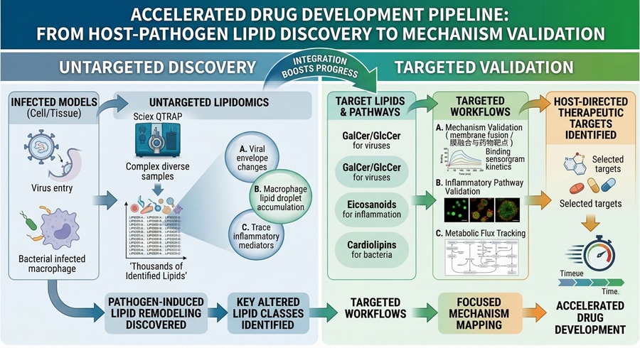
Host Glycosphingolipids Essential for Viral Membrane Fusion

Method used

To understand how enveloped bandaviruses breach host cells, researchers utilized targeted host-pathogen lipidomics to quantify intracellular GalCer and GlcCer levels in 293T cells following pharmacological inhibition. This was coupled with Surface Plasmon Resonance (SPR) assays to evaluate HRTV Gc binding kinetics to specifically formulated liposomes.

Result obtained

The study directly proved that host glucosylceramide (GlcCer) is structurally essential for bandavirus glycoprotein-mediated membrane fusion. Depleting host GlcCer shifted lipid availability and effectively abolished viral entry, demonstrating a clear, targetable lipid-dependent infection mechanism.

Recommended path

Discovery Profiling → Targeted Sphingolipid Validation

Heatmap of host-pathogen lipidomics data showing GalCer and GlcCer changes in 293T cells during bandavirus entry analysis.
Heat map of the levels of intracellular GalCer and GlcCer in 293T cells with or without NB-DNJ treatment.
SPR sensorgram showing host membrane lipid binding by HRTV Gc, supporting glucosylceramide-dependent viral fusion.
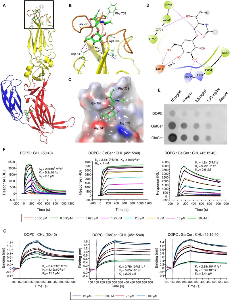
SPR assay of HRTV Gc binding kinetics to the indicated liposomes at neutral pH.
Reference: Knight et al., 2018. Lipid droplet formation in Mycobacterium tuberculosis infected macrophages requires IFN-γ/HIF-1α signaling and supports host defense. PLoS Pathogens. DOI: 10.1371/journal.ppat.1006874
Who needs this: Infection biologists and immunologists studying intracellular bacteria, macrophage lipid droplet dynamics, and infection-induced inflammatory lipid metabolism.

Macrophage Lipid Droplets in Mycobacterium tuberculosis Infection

Method used

The study investigated the M. tuberculosis–macrophage interaction by combining in vitro confocal microscopy of infected macrophages with in vivo histology (Oil Red O tissue staining) to meticulously track neutral lipid accumulation and eicosanoid biology under specific IFN-γ/HIF-1α signaling constraints.

Result obtained

The multi-level profiling successfully linked IFN-γ signaling directly to lipid droplet formation both in isolated cells and within complex in vivo tuberculosis lesions. The data illustrates how host lipid remodeling and localized neutral lipid accumulation support structural defense biology against intracellular bacterial pathogens.

Recommended path

Untargeted Discovery → Targeted Neutral Lipid / Eicosanoid Panel

Confocal microscopy of macrophage lipid droplets during M. tuberculosis infection, highlighting IFN-gamma-dependent host lipid remodeling.
Macrophage LD formation during M. tuberculosis infection requires IFN-γ.
Oil Red O tissue image showing lipid droplet formation in tuberculosis lesions, supporting in vivo host-pathogen lipidomics context.
IFN-γ signaling is required for LD formation during M. tuberculosis infection in vivo.

Frequently Asked Questions (FAQ)

How do you approach differentiating host-derived and pathogen-derived lipids in a co-culture infection model?
Distinguishing cellular origins requires advanced analytical strategies. We utilize highly curated mass spectral databases to identify species generally unique to the pathogen (e.g., bacterial ornithinolipids) versus the mammalian host. In highly complex overlaps, differential expression analysis against pure host and pure pathogen controls provides the statistical foundation for assigning lipid origin.
Can you quantify low-abundance eicosanoids and oxylipins in infected tissue samples?
Yes. Eicosanoids orchestrate the host inflammatory response but are easily masked by highly abundant structural lipids. Our eicosanoid and oxylipin panels use targeted LC-MS/MS coupled with Solid-Phase Extraction (SPE) to selectively concentrate these inflammatory lipid mediators, allowing us to map the inflammatory burst even in minute tissue biopsies or bronchoalveolar lavage fluid (BALF).
What are the biosafety requirements for submitting samples infected with viable pathogens?
We cannot accept live, infectious BSL-3 or highly pathogenic BSL-2 samples. Instead, we provide validated, solvent-based inactivation protocols (e.g., cold methanol/chloroform extractions). Your team performs this inactivation safely within your own biosafety cabinet. Once denatured and rendered non-infectious, the stable lipid extracts can be safely shipped to us on standard dry ice.
How does lipidomics help identify host pathways hijacked by viruses for envelope assembly?
By utilizing targeted and untargeted lipidomics, we profile purified viral particles and compare them directly against the uninfected host cell membrane. If the virus is significantly enriched in a specific host lipid class (such as cholesterol or a specific ceramide), it highlights exactly which host metabolic pathway the virus requires for successful budding and cellular entry.
Why is targeted validation necessary after an untargeted lipidomics discovery phase?
Untargeted lipidomics is powerful for generating hypotheses and observing broad infection lipid metabolism trends. However, targeted validation utilizes specific internal standards and optimized chromatographic gradients to absolutely quantify the lipids of interest. This step is critical for confirming biomarker reliability, satisfying peer-review standards, and comparing exact drug efficacies.
Is spatial lipidomics (MALDI-MSI) applicable to analyzing infected tissue lesions, like granulomas?
Yes. MALDI-Imaging Lipidomics allows us to take a frozen slice of an infected tissue and visually map the distribution of specific lipid species directly across the histological landscape. This preserves the spatial context, helping researchers see exactly where lipid remodeling is occurring relative to the necrotic core or the invading pathogen colony.
How do you prevent the ex vivo degradation of sensitive inflammatory lipids during sample collection?
Inflammatory lipids degrade rapidly. We provide standard operating procedures (SOPs) for immediate flash freezing in liquid nitrogen exactly at the point of biological collection. The samples must then be extracted using deeply chilled solvents pre-loaded with antioxidant shields to instantly halt endogenous lipase activity and prevent auto-oxidation.
Do you support multi-omics integration for infection models?
Yes. To fully elucidate how a pathogen manipulates host metabolism, we can assist in mathematically aligning your host/pathogen transcriptomic datasets (RNA-Seq) with our quantitative lipidomic shifts. This provides comprehensive mapping that establishes a clear pathway from gene expression to physical membrane remodeling.
* Our services can only be used for research purposes and Not for clinical use.

Applications:


Online Inquiry

CONTACT US

Copyright © 2026 Creative Proteomics. All rights reserved.