Our services have earned the trust of companies, schools, and organizations globally, and we remain dedicated to maintaining that trust.
What Are Isoprostanes?
Isoprostanes are prostaglandin-like compounds formed by free radical-catalyzed peroxidation of arachidonic acid esterified in membrane phospholipids. Unlike prostaglandins — which are produced enzymatically by cyclooxygenases (COX-1/COX-2) — isoprostanes are generated non-enzymatically at the site of lipid peroxidation, released from membranes by phospholipase A2, and excreted in urine. This non-enzymatic origin makes them specific reporters of oxidative stress rather than inflammation.
The most widely measured isoprostane is 8-iso-PGF2α (also called 15-F2t-isoprostane or 8-isoprostane). Elevated urinary 8-iso-PGF2α is associated with cardiovascular disease, diabetes, neurodegenerative disorders, and environmental exposures. Levels decrease with smoking cessation, aerobic exercise, and antioxidant therapy — making isoprostanes an objective pharmacodynamic endpoint in intervention studies. However, accurate measurement requires LC-MS/MS: ELISA-based kits cross-react with prostaglandin isomers and can overestimate concentrations by 2- to 10-fold.
Beyond F2-isoprostanes derived from arachidonic acid, F4-neuroprostanes are formed from DHA — the most abundant polyunsaturated fatty acid in brain grey matter — and provide a brain-specific readout of neuronal oxidative damage. Isofurans, produced under elevated oxygen tension, complement isoprostanes when tissue oxygenation is a confounding variable.
Isoprostane Analysis Service in Creative Proteomics
F2-isoprostanes, F4-neuroprostanes, and isofurans are quantified by isotope-dilution LC-MS/MS with deuterated internal standards (8-iso-PGF2α-d4, 8-iso-PGE2-d4). Samples are collected and processed under strict antioxidant stabilization — BHT, EDTA, and N-ethylmaleimide are added at collection to prevent artifactual isoprostane formation ex vivo. The method achieves an LLOQ of 0.01 ng/mL for 8-iso-PGF2α in plasma, with isomer baseline separation on UPLC C18 (1.7 μm) confirmed by ion-ratio verification across 3 MRM transitions per analyte.
F2-Isoprostane Panel (8 Analytes)
8-iso-PGF2α, 8-iso-PGE2, iPF2α-VI, iPF2α-III, 2,3-dinor-8-iso-PGF2α (urinary metabolite), 8-iso-15(R)-PGF2α, 5-F2t-isoprostane, 15-F2t-isoprostane. Each quantified against its deuterated internal standard at LLOQ 0.01 ng/mL.
F4-Neuroprostane Panel (DHA-Derived)
F4-neuroprostanes are formed from DHA peroxidation in neuronal membranes — they are the only lipid peroxidation markers specific to brain grey matter. Quantified in CSF, brain tissue, and plasma. Essential for Alzheimer's, Parkinson's, and traumatic brain injury studies where CNS-specific oxidative damage readouts are required.
Isofuran Panel
Isofurans are preferentially formed over isoprostanes under elevated oxygen tension — when pO2 is high, the isoprostane pathway is suppressed and isofuran formation dominates. Quantifying both isoprostanes and isofurans provides an oxygenation-independent assessment of lipid peroxidation, critical for hyperoxia studies and tissue oxygenation confounders.
Stabilized Sample Collection
BHT (radical scavenger), EDTA (metal chelator), and N-ethylmaleimide (thiol alkylator) are added at the point of collection to prevent artifactual isoprostane formation. Without stabilization, 8-iso-PGF2α can increase 5- to 20-fold ex vivo within 30 minutes — the single largest source of error in published isoprostane data.
Complementary Lipid Peroxidation Markers
Extend your isoprostane panel with malondialdehyde (MDA), 4-hydroxynonenal (4-HNE), 4-hydroxyhexenal (4-HHE), and oxysterols (7-KC, 7β-HC) for multi-pathway oxidative stress profiling from the same sample. Each marker is quantified by a validated, stabilized method.
Isoprostane Detection Panel
- Isoprostane Panel
- Related Services
Isoprostane & Related Lipid Peroxidation Markers
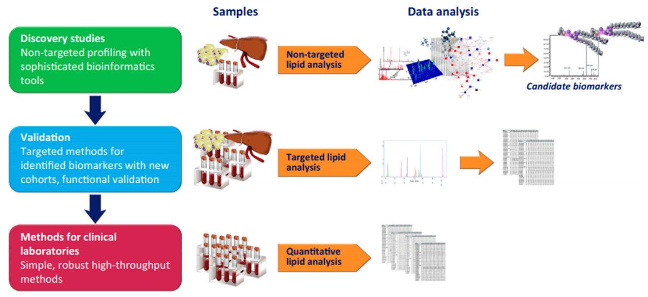
Isotope-dilution LC-MS/MS with deuterated IS. 3 MRM transitions per analyte with ion-ratio confirmation. LLOQ 0.01 ng/mL for F2-isoprostanes. Sample stabilization with BHT/EDTA/NEM at collection.
| Analyte | Precursor | IS | LLOQ | Key Application |
|---|
| 8-iso-PGF2α (15-F2t-IsoP) | Arachidonic acid (F2-IsoP) | 8-iso-PGF2α-d4 | 0.01 ng/mL | Gold-standard systemic oxidative stress biomarker; cardiovascular, metabolic, and neurodegenerative disease |
| 8-iso-PGE2 | Arachidonic acid (F2-IsoP) | 8-iso-PGE2-d4 | 0.02 ng/mL | Vasoconstrictive isoprostane; elevated in hypertension and preeclampsia |
| iPF2α-VI | Arachidonic acid (F2-IsoP) | 8-iso-PGF2α-d4 | 0.02 ng/mL | Second most abundant F2-isoprostane; independently associated with coronary artery disease |
| 2,3-dinor-8-iso-PGF2α | 8-iso-PGF2α metabolite | 2,3-dinor-8-iso-PGF2α-d4 | 0.03 ng/mL | Urinary β-oxidation metabolite; reflects systemic rather than renal isoprostane production |
| Additional F2-Isoprostanes | Arachidonic acid (F2-IsoP) | 8-iso-PGF2α-d4 | 0.02–0.05 ng/mL | iPF2α-III, 8-iso-15(R)-PGF2α, 5-F2t-isoprostane, 15-F2t-isoprostane — included in the F2-isoprostane panel for comprehensive regioisomer coverage |
| F4-Neuroprostanes | DHA (F4-NeuroP) | NeuroP-d4 | 0.05 ng/mL | Brain-specific neuronal oxidative damage; Alzheimer's, Parkinson's, TBI |
| Isofurans | Arachidonic acid | 8-iso-PGF2α-d4 | 0.03 ng/mL | Complement to isoprostanes under elevated pO2; hyperoxia and ventilation studies |
| MDA (Malondialdehyde) | PUFA peroxidation | MDA-d2 | 0.05 ng/mL | Broad-spectrum lipid peroxidation endpoint; TBARS assay alternative |
| 4-HNE / 4-HHE | ω-6 / ω-3 PUFA | 4-HNE-d3 | 0.05 ng/mL | Reactive aldehydes; protein adduct formation, ferroptosis, and cellular stress signaling |
Why LC-MS/MS — Not ELISA — for Isoprostane Quantification
- ELISA cross-reactivity overestimates isoprostane concentrations 2- to 10-fold. LC-MS/MS with isomer baseline separation and 3 MRM transitions per analyte provides unambiguous identification and quantification.
- Deuterated internal standards (8-iso-PGF2α-d4, 8-iso-PGE2-d4, 2,3-dinor-8-iso-PGF2α-d4) spiked at collection correct for extraction efficiency, matrix effects, and artifactual formation.
- Stabilized collection — BHT + EDTA + NEM added at the point of blood draw. Without this, ex vivo isoprostane formation produces 5- to 20-fold artifacts within 30 minutes at room temperature.
- UPLC C18 (1.7 μm) baseline-resolves 8-iso-PGF2α from PGF2α — isomers that differ only in side-chain stereochemistry and are indistinguishable by ELISA.
- Ion-ratio verification across 3 MRM transitions per analyte — MRM1 for quantification, MRM2/MRM3 for identity confirmation — meets bioanalytical method validation acceptance criteria.
- Dual isoprostane + isofuran quantification provides oxygenation-independent lipid peroxidation assessment — critical when tissue pO2 is a confounding variable.
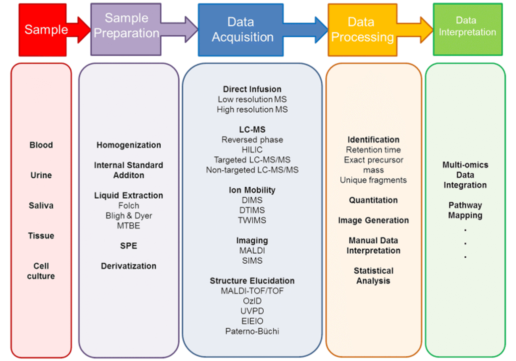
Isoprostane Analysis Workflow & Platform

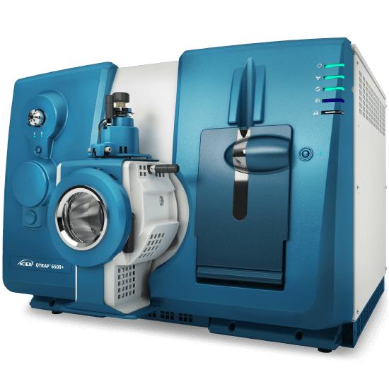
SCIEX Triple Quad 6500+ — scheduled MRM with 3 transitions per analyte and ion-ratio confirmation for isomer-specific quantification.
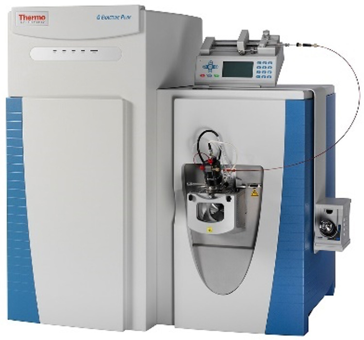
Thermo Q Exactive HF-X Orbitrap — high-resolution confirmation and untargeted screening for novel lipid peroxidation products at 120,000 resolution.
LC-MS/MS Method Specifications
| Parameter | Specification |
|---|
| Platform | SCIEX Triple Quad 6500+ with Turbo V ion source; negative ion ESI |
| Acquisition | Scheduled MRM; 3 transitions per analyte (quantifier + 2 qualifier); ion-ratio verification |
| Chromatography | UPLC C18 (2.1 × 100 mm, 1.7 μm); 15-min gradient; baseline resolution of 8-iso-PGF2α from PGF2α |
| IS | 8-iso-PGF2α-d4, 8-iso-PGE2-d4, 2,3-dinor-8-iso-PGF2α-d4; spiked at collection |
| Stabilization | BHT (0.005%) + EDTA (1 mM) + NEM (1 mM) added at point of sample collection |
| Precision | Intra-assay CV below 8%; inter-assay CV below 12% |
| Calibration | 7-point matrix-matched; 1/x2 weighted; r2 ≥ 0.995 |
Isoprostane Analysis — Results & Data
Isoprostane Quantification Report
Results provided:
- Absolute concentrations (ng/mL or pg/mg) per analyte with deuterated IS recovery per sample
- Urinary isoprostanes normalized to creatinine; F2-isoprostane sum and neuroprostane/isoprostane ratio
- Ion-ratio verification data for isomer identity confirmation; calibration curve metrics; blank carryover
Integrated analytical validation: UPLC baseline separation of 8-iso-PGF2α from PGF2α (panel a), 3-transition ion-ratio identity confirmation (panel b), and stable isotope dilution calibration with r2 ≥ 0.995 (panel c).
Integrated biological results: urinary 8-iso-PGF2α across experimental groups (panel a), multi-marker oxidative lipid damage heatmap (panel b), and oxidative stress interpretation pathway map (panel c).
Differential Analysis
- Group comparisons with FDR-corrected significance per analyte
- Volcano plots and heatmaps; F2-isoprostane sum as composite oxidative stress index
- Isoprostane/isofuran ratio for oxygenation-independent interpretation
Multi-Omics & Pathway Integration
- KEGG arachidonic acid metabolism pathway with fold-change overlay
- Correlation of isoprostanes with complementary lipid peroxidation markers (MDA, 4-HNE, oxysterols)
- Publication-ready figures (TIFF/PDF, 600 dpi)
Explore our Lipidomics Solutions brochure to learn more about isoprostane analysis and our complete lipid peroxidation marker profiling capabilities.
Download Brochure
What Isoprostane Analysis Is Used For
Cardiovascular & Metabolic Disease
8-iso-PGF2α is independently associated with coronary artery disease, hypertension, and post-operative atrial fibrillation. Elevated in diabetes and metabolic syndrome. Used as a pharmacodynamic endpoint in statin, fenofibrate, and antioxidant intervention trials. Levels decrease with smoking cessation, exercise, and weight reduction.
Neurodegeneration & CNS Oxidative Damage
F4-neuroprostanes provide the only brain-specific lipid peroxidation readout — derived from DHA enriched in neuronal membranes. Elevated in Alzheimer's CSF, Parkinson's substantia nigra, and traumatic brain injury. Isofurans complement neuroprostanes when tissue oxygenation is altered.
Antioxidant & Dietary Intervention Studies
Isoprostanes are the preferred objective endpoint for antioxidant efficacy — they decrease in response to vitamin E, polyphenol, and flavonoid interventions. Urinary 2,3-dinor-8-iso-PGF2α reflects systemic production independent of renal contribution.
Environmental & Occupational Exposure
Isoprostanes increase with air pollution (PM2.5, diesel exhaust), cigarette smoke, heavy metals, and radiation exposure. Used as a dosimetry biomarker in environmental health studies and occupational monitoring.
Sample Requirements for Isoprostane Analysis
| Sample Type | Minimum | Critical Handling |
|---|
| Plasma (EDTA) | 50–200 μL | BHT (0.005%) + EDTA (1 mM) + NEM (1 mM) added at collection. Centrifuge immediately at 4°C. Snap-freeze within 15 min. Without stabilization, ex vivo isoprostane formation produces 5- to 20-fold artifacts. |
| Urine | 1–5 mL | Spot or 24 h collection with BHT preservative. Report creatinine for normalization. 2,3-dinor-8-iso-PGF2α in urine reflects systemic production. |
| CSF | 500 μL–1 mL | Polypropylene tube; add BHT/EDTA at collection. CSF isoprostanes and neuroprostanes reflect CNS oxidative damage. |
| Tissue | 10–50 mg | Snap-freeze in LN2 within 30 s. Add BHT/EDTA to homogenization buffer. Tissue isoprostanes measure in situ lipid peroxidation. |
| Cell Culture / Supernatant | 1–5 × 10⁶ cells or 1 mL medium | Wash cells with ice-cold PBS + BHT/EDTA. For supernatant, add BHT immediately after collection. Avoid phenol red — it interferes with SPE extraction. |
FAQ — Isoprostane Analysis
Why use LC-MS/MS instead of ELISA for isoprostane measurement?
ELISA kits cross-react with prostaglandin isomers that co-elute with 8-iso-PGF2α, producing 2- to 10-fold overestimates. LC-MS/MS separates these isomers by UPLC (C18 1.7 μm) and uses 3 MRM transitions per analyte — a quantifier and two qualifiers — with ion-ratio verification for unambiguous identification. This is the consensus method recommended by the Isoprostane Nomenclature Committee.
Why is sample stabilization with BHT/EDTA/NEM critical?
Arachidonic acid in collected blood continues to undergo auto-oxidation ex vivo, generating artifactual isoprostanes within minutes. Plasma left at room temperature for 30 minutes without stabilization can show 5- to 20-fold increases in 8-iso-PGF2α. BHT scavenges free radicals, EDTA chelates metal catalysts, and N-ethylmaleimide alkylates free thiols. All three must be added at the point of collection — not after centrifugation.
What is the difference between F2-isoprostanes and F4-neuroprostanes?
F2-isoprostanes derive from arachidonic acid (C20:4 ω-6), which is ubiquitous in all cell membranes. F4-neuroprostanes derive from DHA (C22:6 ω-3), which is selectively enriched in brain grey matter and retinal photoreceptors. Neuroprostanes provide a CNS-specific oxidative damage readout — useful in Alzheimer's, Parkinson's, TBI, and retinal degeneration studies where systemic isoprostanes may not reflect neuronal damage.
When should I measure isofurans in addition to isoprostanes?
Under normoxic conditions, isoprostane formation dominates. Under elevated pO2 (hyperoxia, mechanical ventilation, reperfusion), the isoprostane pathway is suppressed and isofuran formation increases. Measuring both provides an oxygenation-independent lipid peroxidation index — important for critical care studies, hyperoxia models, and ischemia-reperfusion experiments where tissue pO2 is a variable.
What sample types do you accept for isoprostane analysis?
Plasma (EDTA, 50–200 μL), urine (spot or 24 h, 1–5 mL), CSF (500 μL–1 mL), tissue (10–50 mg, snap-frozen), and cell culture (1–5 × 10⁶ cells or 1 mL medium). All sample types require BHT/EDTA stabilization at the point of collection. Detailed collection protocols are provided with each project to ensure pre-analytical consistency.
Can isoprostanes be analyzed alongside prostaglandins from the same sample?
Yes. Isoprostanes and prostaglandins can be quantified from the same sample extract using separate MRM acquisition methods on the same LC-MS/MS platform. This provides a direct comparison of non-enzymatic (isoprostane) and enzymatic (prostaglandin) lipid peroxidation pathways. Additional sample volume (100–200 μL plasma) is required for the combined panel.