Very Long Chain Fatty Acids (VLCFA) Analysis — LC-MS/MS Quantification of C22:0, C24:0 & C26:0

Very long chain fatty acids (VLCFA, ≥C22) are degraded almost exclusively in peroxisomes — making their accurate quantification the most direct biochemical readout of peroxisomal β-oxidation function. Creative Proteomics delivers targeted LC-MS/MS analysis of docosanoic acid (C22:0), tetracosanoic acid (C24:0), and hexacosanoic acid (C26:0) with calculated C24/C22 and C26/C22 ratios — each analyte quantified against its own deuterated internal standard.

What we measure: C22:0 (behenic acid) · C24:0 (lignoceric acid) · C26:0 (cerotic acid) — plus C24/C22 & C26/C22 ratios

Why it matters: Peroxisomal β-oxidation research · ABCD1/PEX gene models · myelin & skin barrier lipid studies · preclinical drug evaluation

Request Analysis

Submit Your Request Now

×

Our services have earned the trust of companies, schools, and organizations globally, and we remain dedicated to maintaining that trust.

Boston University
University at Buffalo
UMass Medical School
Hawaii Pacific University
Medizinische Fakultät
Louisiana State University
Nyulangone
Nature's Fynd
Dietary Supplement Manufacturer
  • Service We Provide
  • VLCFA Panel & Methods
  • Advantages
  • Workflow
  • Results and Data Analysis
  • Sample Requirements
  • FAQ
  • Publication

What Are Very Long Chain Fatty Acids?

Very long chain fatty acids (VLCFA) are fatty acids with aliphatic carbon chains of 22 or more carbon atoms. The three most studied species are docosanoic acid (C22:0, behenic acid), tetracosanoic acid (C24:0, lignoceric acid), and hexacosanoic acid (C26:0, cerotic acid). Unlike shorter-chain fatty acids oxidized in mitochondria, VLCFA undergo β-oxidation almost exclusively in peroxisomes — making their plasma and tissue concentrations a direct biochemical readout of peroxisomal function.

Beyond their diagnostic value, VLCFA are essential structural components: C22–C26 fatty acids are incorporated into myelin sphingolipids that maintain axonal integrity, and into epidermal acylceramides that form the skin's water-impermeable barrier. Accurate, species-level quantification — rather than total VLCFA measurement — is therefore critical across neurobiology, dermatology, and metabolic research.

VLCFA Service in Creative Proteomics

We provide dedicated LC-MS/MS quantification of very long chain fatty acids using multiple reaction monitoring (MRM) with stable isotope-labeled internal standards (d4-C22:0, d4-C24:0, d4-C26:0). Each analytical batch includes matrix-matched calibration and comprehensive QC reporting.

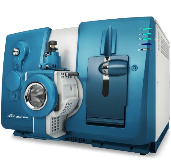
Absolute VLCFA Quantification

LC-MS/MS MRM quantification of C22:0, C24:0, and C26:0 against matched deuterated internal standards on a SCIEX Triple Quad 6500+. LLOQ of 0.2 ng/mL for C22:0 and C24:0, 0.1 ng/mL for C26:0, with linear dynamic range spanning 4 orders of magnitude and intra-batch CV below 10%.

C24/C22 & C26/C22 Ratio Analysis

Automated ratio calculation from the same LC-MS/MS run, normalizing for inter-sample variability in total lipid content. The C26/C22 ratio is the most sensitive single indicator of peroxisomal β-oxidation status, while C24/C22 provides complementary information for peroxisomal biogenesis assessment.

Multi-Matrix VLCFA Profiling

Validated protocols for EDTA plasma (100 μL), serum, tissue homogenates (10–50 mg), cell pellets (1×10⁶ cells), CSF, dried blood spots, and urine. Each matrix uses optimized lipid extraction with BHT antioxidant protection.

Peroxisomal Biology Research Support

Preconfigured VLCFA panels for ABCD1 mutation models, PEX gene knockout models, and D-bifunctional protein deficiency studies. In-house reference data available for C57BL/6 and BALB/c mice, Sprague-Dawley and Wistar rats, and zebrafish.

Advanced Data Analysis & Interpretation

PCA plots, volcano plots for differential VLCFA abundance, correlation heatmaps, and KEGG peroxisomal β-oxidation pathway mapping with fold-change overlay. Machine learning-assisted feature selection available for multi-cohort studies.

VLCFA Detection Panel & LC-MS/MS Method

Three very long chain fatty acid species are quantified using LC-MS/MS MRM with matched deuterated internal standards. Quantification is performed by stable isotope dilution — each VLCFA analyte is measured against its own labeled analog (d4-C22:0, d4-C24:0, d4-C26:0) spiked into the sample before extraction, mathematically correcting for matrix effects, adsorptive losses, and ionization variability in a single step.

AnalyteCommon NameISLLOQCV
Docosanoic Acid (C22:0)Behenic acidd4-C22:00.2 ng/mL3.8%
Tetracosanoic Acid (C24:0)Lignoceric acidd4-C24:00.2 ng/mL4.2%
Hexacosanoic Acid (C26:0)Cerotic acidd4-C26:00.1 ng/mL5.1%

C24/C22 and C26/C22 ratios are calculated from the same LC-MS/MS run. The C26/C22 ratio is the most sensitive biochemical indicator of peroxisomal β-oxidation status; the C24/C22 ratio provides complementary information. Method: SCIEX Triple Quad 6500+ with Scheduled MRM, C18 column (2.1 × 100 mm, 1.7 μm), 12-min ACN → IPA gradient with 0.1% formic acid. Extraction: MTBE-based with 0.005% BHT + 1 mM EDTA under nitrogen. Calibration: 7-point matrix-matched curves (r2 ≥ 0.995, 1/x2 weighted). QC: 3 levels per batch with pooled biological QC for longitudinal studies. GC-MS available as confirmatory platform for isomer resolution.

Related Fatty Acid Services

Pair VLCFA analysis with complementary services using matched sample preparation protocols:

VLCFA Analysis Workflow & Instrument Platform

VLCFA Targeted Lipidomics Workflow — from sample preparation through LC-MS/MS MRM to quantitative report

SCIEX Triple Quad 6500+

SCIEX Triple Quad 6500+ (Figure from SCIEX)

Thermo Scientific UltiMate 3000

Thermo Scientific UltiMate 3000 (Figure from Thermo Fisher)

Agilent 7890A GC System

Agilent 7890A GC System (Figure from Agilent)

Why Choose Our VLCFA Analysis Platform

  • Absolute quantification with matched deuterated internal standards (d4-C22:0, d4-C24:0, d4-C26:0) spiked pre-extraction — correcting for matrix effects, extraction efficiency, and ionization variability in a single step.
  • LLOQ of 0.2 ng/mL (C22:0, C24:0) and 0.1 ng/mL (C26:0) — enabling VLCFA detection in volume-limited samples: dried blood spots, microdialysates, small biopsies.
  • Dual-platform capability — LC-MS/MS as primary (no derivatization, direct VLCFA detection) with GC-MS available for confirmatory isomer resolution.
  • Antioxidant-protected sample preparation: 0.005% BHT + 1 mM EDTA in all extraction solvents, nitrogen-blanketed evaporation, cold-chain at ≤4°C.
  • Transparent QC: calibration r2, IS recovery, blank carryover, pooled QC CV tracking — every metric reported per batch.
  • In-house VLCFA reference data for C57BL/6, BALB/c, Sprague-Dawley, Wistar, and zebrafish — accelerate preclinical interpretation.

VLCFA Analysis — Results and Data Analysis

VLCFA Quantification & Ratio Report

Results we provide:

  • Absolute concentrations for C22:0, C24:0, C26:0 in nmol/mL or nmol/mg
  • Calculated C24/C22 and C26/C22 ratios with batch-level distributions
  • Internal standard recovery per sample
  • PCA for group clustering and outlier identification

QC metrics included:

  • Inter-batch CV, calibration curve r2, blank carryover
  • 3-level QC sample tracking per analytical batch
VLCFA MRM chromatogram overlay — C22:0 C24:0 C26:0 with d4-labeled internal standards

Overlay of MRM chromatograms for C22:0, C24:0, and C26:0 with d4-labeled internal standards — 12-min gradient, baseline resolution.

In-Depth Comparative Analysis

Results provided:

  • Pairwise group comparisons (t-test, ANOVA, non-parametric)
  • Volcano plots — fold-change vs significance per VLCFA species
  • Correlation matrix linking VLCFA to phenotypic or omics variables
  • Heatmap visualization across all individual samples

Pathway & Multi-Omics Integration

Results provided:

  • KEGG peroxisomal β-oxidation pathway with VLCFA fold-change overlay
  • Integration with glycerophospholipid or sphingolipid profiling data
  • ML-assisted feature ranking for VLCFA biomarker identification
  • Publication-ready figures (TIFF/PDF, 600 dpi)

Explore our Lipidomics Solutions brochure to learn more about the full spectrum of VLCFA and targeted lipidomics analysis capabilities.

Download Brochure

What Our VLCFA Analysis Used For

Peroxisomal Disorder Models

Plasma VLCFA profiling is the primary biochemical endpoint for X-ALD (ABCD1 knockout) and Zellweger spectrum (PEX gene knockout) models. C26/C22 ratio serves as a pharmacodynamic biomarker in gene therapy and small-molecule intervention studies.

Myelin & Neurobiology

Myelin is enriched in VLCFA-containing galactocerebrosides (C24:0/C24:1) and sulfatides. Brain regional VLCFA profiling supports leukodystrophy, adrenomyeloneuropathy, and multiple sclerosis research.

Skin Barrier & Ceramide Biology

Epidermal acylceramides require ultra-long C26–C36 ω-hydroxy fatty acids synthesized by ELOVL1 and ELOVL4. VLCFA deficiency in stratum corneum disrupts barrier function — studied in atopic dermatitis, ichthyosis, and reconstructed epidermal models.

Metabolic & Cardiovascular Research

Plasma C24:0 and C26:0-ceramides correlate with insulin resistance, NAFLD/NASH, and cardiovascular risk. Tissue VLCFA profiling in dietary intervention models and genetic obesity models (ob/ob, db/db).

Drug & Gene Therapy Development

VLCFA reduction is a primary endpoint in X-ALD therapies — Lorenzo's oil, bezafibrate, and ABCD1 gene therapy are all monitored via plasma C26:0 and C26/C22 ratio. ELOVL1-targeting agents for skin disorders assessed by acylceramide VLCFA profiling.

Dietary & Nutritional Lipidomics

Dietary VLCFA quantification in peanut oil (C22:0, C24:0), mustard seed oil (C22:1 erucic acid), and marine oils (C20–C22 ω-3 PUFA). VLCFA tissue incorporation and bioavailability in controlled feeding studies, functional food development, and infant formula research.

Sample Requirements for VLCFA Analysis

Sample TypeCollection & ProcessingMinimum QtyCritical Notes
Plasma (EDTA)Lavender-top EDTA tube; centrifuge within 30 min at 4°C, 2,000×g, 10 min100 μLEDTA plasma preferred over serum — platelet VLCFA release during clotting. Aliquot immediately; snap-freeze in LN2; store -80°C. No freeze-thaw.
TissueRinse with ice-cold PBS; snap-freeze in LN2 within 5 min10–50 mgPeroxisome-rich tissues (liver, adrenal) degrade fastest. For brain: dissect on ice-cold stage. For skin: remove subcutaneous fat.
Cell PelletsWash 2× ice-cold PBS; pellet 300×g, 5 min, 4°C1–5 × 10⁶ cellsWash thoroughly to remove medium lipids. Normalize to cell count or total protein (BCA).
Dried Blood SpotSpot onto Whatman 903 card; dry 3–4 h at RT1 × 3.2 mm punchValidated for mouse tail-vein studies. Store with desiccant at -20°C (short) or -80°C (long). Avoid humidity.
CSFPolypropylene tube; centrifuge within 30 min, 4°C500 μL–1 mLVLCFA abundance in CSF is very low — use polypropylene tubes to reduce adsorptive loss. Contact us for micro-scale protocol.

FAQ — VLCFA Analysis

What is the detection limit for VLCFA analysis?

Our LC-MS/MS MRM method achieves LLOQ of 0.2 ng/mL for C22:0 and C24:0, and 0.1 ng/mL for C26:0 in plasma — 10–50× more sensitive than conventional GC-MS. This enables VLCFA quantification in volume-limited samples including dried blood spots, microdialysates, and CSF.

How are C24/C22 and C26/C22 ratios calculated and why do they matter?

Ratios are derived from concentrations measured in the same LC-MS/MS run. The C26/C22 ratio is the most sensitive single indicator of peroxisomal β-oxidation status. Ratio-based analysis normalizes for inter-sample variability in total lipid content, providing higher sensitivity than absolute concentrations alone.

How should I prepare and ship samples for VLCFA analysis?

Use EDTA plasma (not serum). Centrifuge within 30 min at 4°C. Aliquot into polypropylene cryovials, snap-freeze in LN2, store at -80°C. Ship on ≥5 kg dry ice. Avoid freeze-thaw — VLCFA recovery drops 5–15% per cycle. Tissues must be snap-frozen within 5 min.

How does LC-MS/MS compare to GC-MS for VLCFA quantification?

LC-MS/MS is our primary platform: direct VLCFA detection without derivatization, 10–50× lower LLOQ, 12-min gradient vs 30–45 min for GC-MS. GC-MS is available as a confirmatory platform for isomer resolution when needed.

How do you prevent VLCFA oxidation during sample preparation?

0.005% BHT + 1 mM EDTA in all extraction solvents, nitrogen-blanketed atmosphere, glass vials for post-extraction steps, cold-chain at ≤4°C, and deuterated IS spiked pre-extraction to mathematically correct for recovery variability.

Do you have reference data for preclinical VLCFA models?

Yes. In-house profiles for C57BL/6, BALB/c (including ABCD1-KO, PEX mutants), Sprague-Dawley, Wistar, and zebrafish. For non-standard models, include 5 wild-type controls for study-specific baselines.

Can raw data be exported for independent re-analysis?

Yes — .mzML and .wiff files with retention times, peak widths, and S/N ratios. Compatible with Skyline, SCIEX OS, Progenesis QI, and XCMS. Methods appendix included for manuscript supplements.

Is this service for clinical diagnostic use?

No. Our VLCFA analysis is for research use only (RUO) and is not CLIA-certified or CAP-accredited. It should not be used for clinical diagnostic or patient management. For clinical VLCFA testing, please contact a certified clinical laboratory. Our service supports preclinical research — peroxisomal biology studies, drug development, biomarker discovery, and mechanistic investigation in model systems.

How many biological replicates do you recommend for VLCFA studies?

A minimum of 5 biological replicates per experimental group for animal studies, and 3 independent culture experiments for cell-based work. Include matched control samples (wild-type or vehicle-treated) processed identically. Technical replicates (2–3 injections from the same extract) are run automatically. For clinical cohort studies, consult our team during study design.

How do I get started with submitting samples?

Click "Request Analysis" or contact us through the inquiry form. Our team will provide a study design consultation, a formal quotation, sample submission guidelines for your matrix, and shipping instructions. Pilot batches are welcome — no minimum sample count.

Publications

References

  1. Moser, H.W., et al. "Plasma very long chain fatty acids in 3,000 peroxisome disease patients and 29,000 controls." Annals of Neurology (1999). https://doi.org/10.1002/1531-8249(199901)45:1<100::AID-ART16>3.0.CO;2-U
  2. Kemp, S., et al. "X-linked adrenoleukodystrophy: clinical, metabolic, genetic and pathophysiological aspects." BBA — Molecular Basis of Disease (2012). https://doi.org/10.1016/j.bbadis.2012.03.012
  3. Valianpour, F., et al. "Analysis of very long-chain fatty acids using electrospray ionization mass spectrometry." Molecular Genetics and Metabolism (2003). https://doi.org/10.1016/S1096-7192(03)00098-2
  4. Kihara, A. "Very long-chain fatty acids: elongation, physiology and related disorders." The Journal of Biochemistry (2012). https://doi.org/10.1093/jb/mvs105
* Our services can only be used for research purposes and Not for clinical use.

Resource:

Platform:


Online Inquiry

CONTACT US

Copyright © 2026 Creative Proteomics. All rights reserved.