Pharmacodynamic & Pharmacokinetic Lipidomics for Lead Optimization

Lipid readouts can translate drug exposure into measurable biology—especially when conventional PD markers are indirect, delayed, or tissue-limited. Our pharmacodynamic lipidomics and pharmacokinetic lipid biomarkers workflows help biopharma teams connect mechanism-driven lipid pathway changes to dose, time, and exposure in discovery and lead optimization. We support plasma lipidomics, cell models, and tissues with fit-for-purpose untargeted profiling, targeted lipidomics quantification, lipid mediator profiling, flux, and spatial lipid mapping.

Key capabilities

  • Exposure–response lipid signatures in plasma lipidomics for PD/PK alignment across dose and time
  • Quantitative confirmation with targeted lipidomics quantification (class panels, internal standards, calibration)
  • Mechanistic deepening via lipid mediator profiling, pathway flux, and MALDI imaging lipidomics in tissues
Request Analysis

Submit Your Request Now

×

Our services have earned the trust of companies, schools, and organizations globally, and we remain dedicated to maintaining that trust.

Boston University
University at Buffalo
UMass Medical School
Hawaii Pacific University
Medizinische Fakultät
Louisiana State University
Nyulangone
Nature's Fynd
Dietary Supplement Manufacturer
  • Trends & Challenges
  • Integrated Solutions
  • Technical Advantages
  • Case Studies
  • FAQ

Solutions For PD/PK Lipidomics

PK/PD lipidomics decisions are rarely linear; they are triggered by study friction—the need for repeatable, exposure-aligned, and decision-ready readouts. We help programs navigate this complexity through an evidence-driven practical loop:

Establish & Stabilize: Start with plasma/serum profiling to identify dose-responsive signatures. If signals appear, transition to targeted lipidomics for absolute quantification and cross-batch comparability.

Refine & Bridge: If signals are inconsistent, resolve pre-analytical noise (stability/batch effects) before expanding cohorts. If systemic PD fails to explain efficacy, bridge into tissue-level analysis to confirm site-of-action biology.

Resolve Deep Mechanism: When causality or localization is required:

  • Oxylipin Profiling: To capture the signaling layer (COX/LOX/CYP) if inflammation is suspected.
  • MALDI Imaging: To map spatial lipid remodeling at the tumor-stroma interface.
  • Metabolic Flux: To definitively distinguish between lipid synthesis and turnover.

PD Signal Not Tracking Dose/Time

Situation: Plasma readouts are subtle or inconsistent across post-dose timepoints; treated vs control separation is near baseline variability, or directionality flips across adjacent timepoints.

Goal: Identify exposure-aligned lipid pathways and convert them into quant-stable PD markers suitable for confirmation and longitudinal tracking.

Recommended Path: Bundle A → Bundle B

Recommended Services:

What You Will Get: Exposure-aligned lipid signature candidates plus a clear conversion path into targeted panels that are more stable across time, dose, and batches.

Need Minimally Invasive, Repeatable PD/PK Marker

Situation: Lead optimization requires repeat sampling across cohorts and dose levels; plasma/serum must support longitudinal PD/PK tracking.

Goal: Establish a minimally invasive lipid biomarker set and translate it into targeted panels for repeatable monitoring (RUO).

Recommended Path: Bundle A → Bundle B

Recommended Services:

What You Will Get: A plasma/serum PD/PK marker strategy built around targeted panels that are easier to replicate across cohorts and timepoints.

Cross-Batch Or Cross-Site Reproducibility Failure

Situation: PD signals don't replicate across batches/sites; effect sizes don't match exposure; likely pathway-specific markers are being diluted by broad profiling or matrix/panel mismatch.

Goal: Rebuild a reproducible PD readout with panel-first endpoints that survive site/batch variation.

Recommended Path: Bundle B (panel-first) → optional Bundle A (context)

Recommended Services:

What You Will Get: A narrowed, reproducibility-first PD panel set with pathway specificity (sphingolipid/sterol modules) that is more stable across sites and batches.

Membrane Remodeling Hypothesis Needs Decision-Grade Quant

Situation: Evidence suggests membrane remodeling (PC/PE/PI, DAG/TAG shifts, ratios), but discovery-level changes aren't robust enough for lead ranking across dose/time/batch.

Goal: Quantify remodeling endpoints with higher confidence and mechanistic specificity to support PD interpretation and optimization decisions.

Recommended Path: Bundle B

Recommended Services:

What You Will Get: Quant-stable membrane remodeling readouts (phospholipid + neutral lipid modules) plus energy/β-oxidation context when acylcarnitines are relevant.

Suspected COX/LOX/CYP Signaling Or Inflammatory Mediators

Situation: Safety pharmacology or efficacy phenotype suggests inflammation signaling; you need mediator-layer PD rather than bulk membrane lipids alone.

Goal: Quantify mediators and connect changes to COX/LOX/CYP pathway branches to support PD biology and risk interpretation.

Recommended Path: Bundle B (mediators)

Recommended Services:

What You Will Get: A mediator-centric PD package (oxylipins/eicosanoids/PGs) that is closer to inflammatory mechanism than bulk lipid classes, with optional upstream lipid drivers.

Plasma PD Moves But Efficacy Does Not Correlate

Situation: Plasma biomarkers move with treatment, but efficacy doesn't correlate; tissue localization, compartment effects, or mitochondrial involvement is suspected.

Goal: Anchor lipid changes to the site of action and identify which lipid modules best reflect pharmacology in tissue.

Recommended Path: Bundle A (tissue bridge) → Bundle B

Recommended Services:

What You Will Get: Tissue-anchored PD interpretation with mitochondrial/membrane modules that better reflect target engagement at the site of action.

Bulk Tissue Averages Hide Localized Drug Response

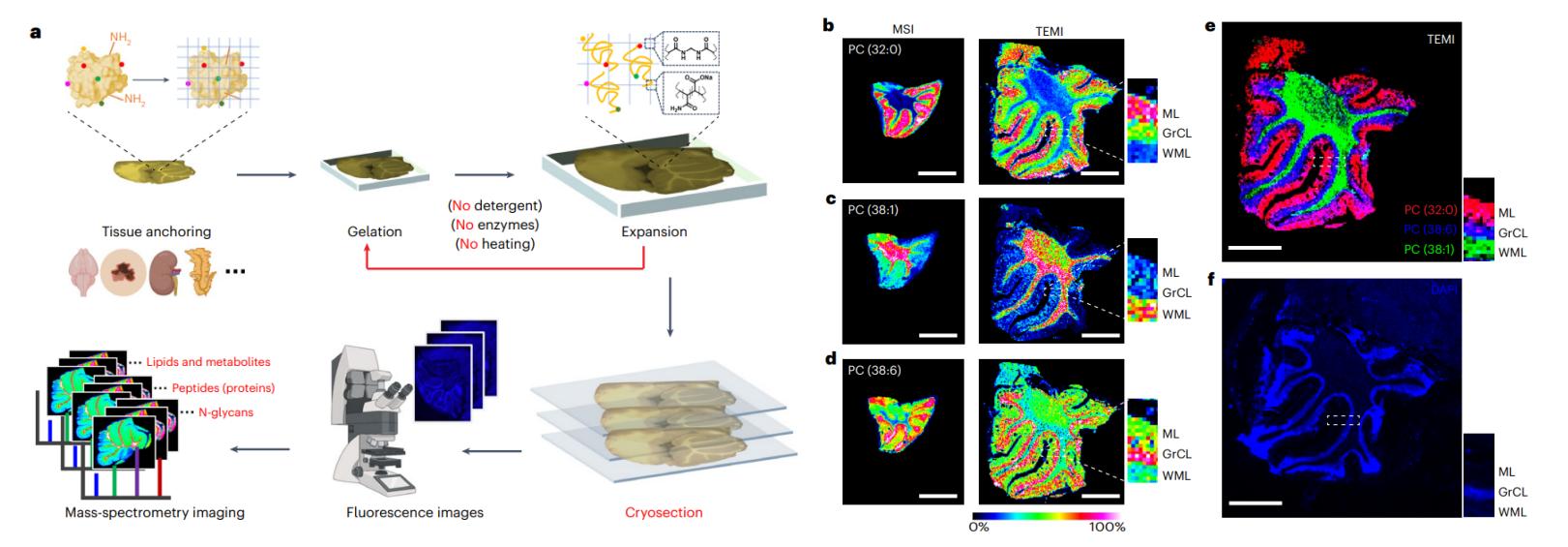
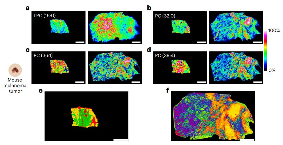
Situation: Heterogeneous tissues dilute localized lipid responses; bulk tissue appears "flat" despite suspected regional pharmacology.

Goal: Visualize spatial lipid changes and quantify region-level differences that explain responder/non-responder patterns or microenvironment effects.

Recommended Path: Bundle C (imaging)

Recommended Services:

What You Will Get: Spatial lipid maps plus region-stratified marker candidates that explain localized PD even when bulk averages fail.

Need Flux Evidence Beyond Steady-State Abundance

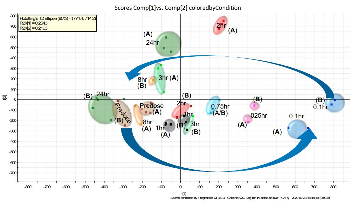
Situation: Steady-state lipid shifts have multiple interpretations; decision-makers require stronger causal evidence (synthesis vs turnover) for MoA/target engagement.

Goal: Provide flux-informed evidence that clarifies whether exposure changes lipid production, remodeling, or turnover.

Recommended Path: Bundle C (flux) → Bundle B

Recommended Services:

What You Will Get: Flux-backed pathway interpretation that separates synthesis vs turnover, supporting stronger PD/PK biomarker decisions at program inflection points.

Case Studies

Publication: Zhang et al., 2025, Nature Methods. https://doi.org/10.1038/s41592-025-02664-9
Who Needs This: Teams whose PD biology is spatially restricted (tumor margin, brain layers, mixed immune infiltration) and cannot be resolved by bulk tissue lipidomics.

Case Study: Single-Cell Spatial Lipid Mapping In Tissue Microenvironments

Method Used

Tissue-expansion MALDI mass-spectrometry imaging (TEMI) to map hundreds of biomolecules including lipids with cellular-scale resolution across mammalian tissues and tumors.

Result Obtained

Higher spatial resolution revealed metabolic heterogeneity and region-specific lipid patterns that were less distinct in unexpanded controls.

Recommended Path

A→C (broad profiling then spatial confirmation).

MALDI imaging lipid maps across cerebellar layers showing layer-enriched phospholipids and an overlay visualization aligned to tissue structure.
TEMI-enabled MALDI imaging maps layer-enriched phospholipids across cerebellar regions (molecular layer, white matter layer, granule cell layer), including single-species ion images and an overlay aligned to DAPI.
Side-by-side lipid ion images of tumor tissue before and after TEMI expansion with clustering segmentation, revealing increased spatial resolution and metabolic heterogeneity.
Comparison of conventional MSI and TEMI-expanded tumor imaging for multiple lipid species (e.g., LPC and PCs), plus clustering-based segmentation showing substantially finer regional heterogeneity after expansion.
Publication: Plumb et al., 2024, Scientific Reports. https://doi.org/10.1038/s41598-024-66764-w
Who Needs This: DMPK and pharmacology teams looking for pharmacokinetic lipid biomarkers that change with exposure in plasma when tissue PD sampling is limited.

Case Study: Pharmacodynamic Plasma Lipids Track Exposure After Small-Molecule Dosing

Method Used

Untargeted LC–MS analysis of mouse plasma after IV and oral gefitinib; multivariate statistics across dense time points; annotation supported by databases and standards where feasible.

Result Obtained

Multiple lipid classes (e.g., PI, PC, LPC, PE, TG) showed time-resolved shifts with rapid onset and recovery trends; the paper discusses a pharmacolipidodynamic relationship between exposure and lipid abundance.

Recommended Path

A→B (discover in plasma, then confirm with quant panels).

PCA scores plot of mouse plasma lipidomics after IV gefitinib, showing early divergence from predose and gradual return toward baseline by 24 hours.
PCA of negative-ion UHPLC–MS plasma lipids across IV gefitinib time points (predose through 24 h), showing a clear time-dependent trajectory and return toward baseline.
Line chart comparing PI(40:5) plasma lipid response with gefitinib PK curve after oral dosing, highlighting exposure-linked lipid dynamics.
Time-resolved change in PI(40:5) after oral gefitinib is shown alongside the gefitinib pharmacokinetic profile, illustrating a PK-linked lipid response pattern.

FAQ

When should lipidomics be used in lead optimization?
Lipidomics is especially useful when conventional PD markers are indirect, delayed, or difficult to measure repeatedly. It helps link drug exposure to pathway-level biology and identify exposure-responsive lipid biomarkers that support target engagement, compound ranking, and translational decision-making.
What sample types and handling practices are best for PD/PK lipidomics?
EDTA plasma is often preferred for longitudinal PD/PK studies because it reduces clotting-related variability and supports repeat sampling across dose groups and time points. Matched predose and multiple post-dose samples are important for exposure–response analysis. To preserve labile lipids, samples should be aliquoted promptly, kept under a strict cold chain, and exposed to as few freeze–thaw cycles as possible. For lipid mediator studies such as oxylipin profiling, minimizing platelet activation during collection is especially important.
What are typical replicate and sample volume requirements?
For discovery-phase studies, at least five biological replicates per group is often a practical starting point, although final study design should reflect model variability and sampling goals. Plasma lipidomics can often be performed from small input volumes, typically around 20–50 µL. For tissues, 10–30 mg is commonly suitable, while spatial imaging usually requires intact fresh- or flash-frozen sections.
Should I start with untargeted profiling or targeted quantification?
Untargeted lipidomics is generally used first when relevant pathways are not yet known, helping identify exposure-responsive lipid signatures across broad classes. Targeted quantification is then used to confirm selected markers with higher quantitative confidence and better cross-batch comparability. In lead optimization, discovery followed by targeted panel development is often the most effective workflow.
How many time points are needed for PK/PD alignment?
Lipid responses can appear quickly and return toward baseline within a short window. For this reason, studies usually benefit from matched predose samples and multiple post-dose time points covering peak exposure and recovery. Dense early sampling is often important when lipid effects are transient.
Can lipidomics support minimally invasive PD biomarker development?
Yes. Plasma or serum lipidomics supports repeatable, minimally invasive sampling, making it well suited for longitudinal PK/PD studies. Once exposure-responsive markers are identified, they can be translated into targeted panels for more consistent monitoring across cohorts, doses, and time points.
How is reproducibility maintained across batches or study sites?
Reproducibility is improved through standardized collection and storage, controlled freeze–thaw exposure, matrix-appropriate internal standards, and targeted confirmation when quantitative comparability is required. When broad profiling is too variable for cross-site studies, pathway-focused targeted panels are often a more robust option.
What if plasma lipid changes do not correlate with efficacy?
When plasma signals track treatment but do not explain efficacy, tissue lipidomics can help determine whether the biologically relevant response is localized at the site of action. Tissue profiling, mitochondrial lipid analysis, or spatial lipidomics can clarify compartment-specific lipid remodeling that may not be visible in circulation.
Can lipidomics reveal spatial heterogeneity or provide stronger mechanistic evidence?
Yes. When bulk tissue averages mask localized pharmacology, MALDI imaging lipidomics can map lipid distributions directly within tissue architecture. When the key question is whether a drug changes lipid synthesis, remodeling, or turnover, metabolic flux analysis provides stronger mechanistic evidence than steady-state abundance alone. Together, these approaches support higher-confidence interpretation of target engagement and MoA.
What information is needed before starting a PD/PK lipidomics study?
Useful starting information includes the compound background, study objective, model system, dose design, sampling schedule, matrix type, and whether the priority is discovery, quantitative confirmation, or translational biomarker development. Existing PK, efficacy, or pathway data can further refine the study strategy.
* Our services can only be used for research purposes and Not for clinical use.

Applications:


Online Inquiry

CONTACT US

Copyright © 2026 Creative Proteomics. All rights reserved.