Our services have earned the trust of companies, schools, and organizations globally, and we remain dedicated to maintaining that trust.
What Are Medium Chain and Long Chain Fatty Acids?
Fatty acids are classified by carbon chain length because chain length determines metabolic fate. Medium chain fatty acids (MCFA, C6–C12) are absorbed directly into the portal vein and transported to the liver, where they enter mitochondria independently of the carnitine shuttle for rapid β-oxidation — making them preferential energy substrates. Long chain fatty acids (LCFA, C13–C21) are re-esterified into triglycerides within enterocytes, packaged into chylomicrons, transported via the lymphatic system, and released into circulation — their cellular uptake and mitochondrial oxidation depend on carnitine palmitoyltransferase (CPT1).
This fundamental metabolic difference means MCFA and LCFA play distinct — and often opposing — roles in health and disease. MCFA promote ketogenesis and are studied in ketogenic diet therapies and infant nutrition. LCFA, particularly saturated palmitic acid (C16:0) and polyunsaturated arachidonic acid (C20:4n6) and DHA (C22:6n3), are central to membrane phospholipid composition, inflammatory signaling, and metabolic disease progression. Accurate, simultaneous quantification of both MCFA and LCFA in a single sample provides a complete picture of fatty acid metabolism — but their different physical properties make this technically demanding.
Why MCFA & LCFA Analysis Is Technically Challenging — and How We Solve It
Accurately quantifying 40+ MCFA and LCFA species across a 4-6 order-of-magnitude concentration range in a single workflow requires methods that specifically address the physicochemical differences between medium- and long-chain analytes. Below are the four principal technical obstacles and the validated approaches used to address each.
| Challenge | Why It Matters | Our Solution |
|---|
| MCFA evaporative loss during sample concentration | C6:0–C10:0 are volatile — up to 50% can be lost during standard nitrogen blow-down, producing falsely low MCFA concentrations and skewed MCFA/LCFA ratios. | Low-temperature (30°C) nitrogen evaporation with trimethylamine co-solvent to reduce vapor pressure. d3-C8:0 and d3-C10:0 internal standards compensate for residual loss. |
| LCFA adsorption to plastic consumables | C16:0 and C18:0 adsorb strongly to polypropylene and polystyrene surfaces at ng/mL concentrations — recoveries can drop below 50% in low-concentration samples. | All post-extraction handling in silanized glass vials with 0.1% formic acid in solvent to compete with surface binding sites. Recovery monitored via IS per sample. |
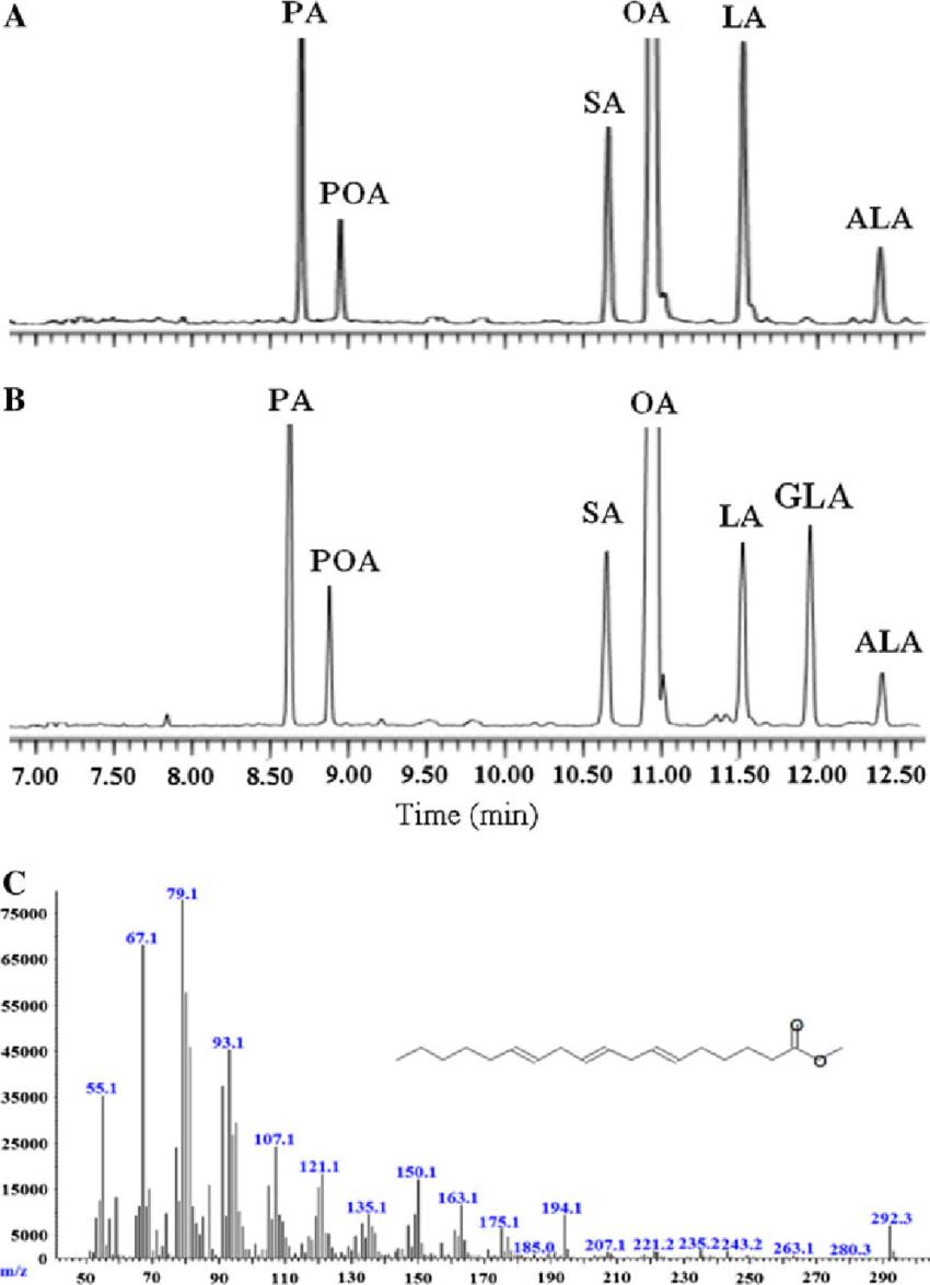
| Isomer separation (C18:1n9 vs C18:1n7; C18:3n3 vs C18:3n6) | Positional and geometric isomers have identical molecular weights and similar fragmentation — standard C18 columns cannot resolve them. Misidentification leads to incorrect biological interpretation. | GC-MS: 60-m highly polar bis-cyanopropyl column (DB-225, 0.25 mm ID, 0.20 μm film) achieves baseline resolution of C18:1n9/n7, C18:2n6/tt, and C18:3n3/n6. Identity confirmed by retention index matching against authenticated standards. |
| Concentration range spanning 4-6 orders of magnitude | In plasma, palmitic acid (C16:0) may be present at mM levels while γ-linolenic acid (C18:3n6) is at sub-μM — both must be quantified from the same injection. | Split-dilution strategy: 1:10 dilution for abundant saturated LCFA (C16:0, C18:0, C18:1) and undiluted analysis for trace PUFA. Both dilutions use identical IS concentrations. Cross-dilution concordance verified (<15% deviation). |
MCFA & LCFA Analysis Service in Creative Proteomics
We offer validated GC-MS and LC-MS/MS quantification of 40+ medium chain and long chain fatty acids. Choose the combined MCFA+LCFA panel for full fatty acid profiling, or select MCFA-only or LCFA-only panels tailored to your research question. Each panel includes stable isotope-labeled internal standards, matrix-matched calibration, and full QC documentation.
Combined MCFA + LCFA Panel (40+ Analytes)
Full fatty acid profiling from C6:0 to C21:0 in a single sample submission — saturated, monounsaturated, and polyunsaturated species. GC-MS primary with LC-MS/MS confirmation for selected analytes. Ideal for untargeted metabolic phenotyping and multi-pathway studies.
MCFA-Only Panel (C6:0–C12:0)
Optimized for volatile MCFA quantification using low-temperature GC-MS with deuterated internal standards (d3-C8:0, d3-C10:0, d3-C12:0). Tailored for ketogenic diet research, MCT oil characterization, infant formula lipid analysis, and sport nutrition studies.
LCFA-Only Panel (C14:0–C21:0)
Focused long chain fatty acid profiling with emphasis on PUFA isomer resolution (ω-3, ω-6, ω-9 families). Applications in cardiovascular research, NAFLD/NASH, inflammation, and membrane lipid remodeling studies. ω-3/ω-6 ratio included.
Dual-Platform Cross-Validation (GC-MS + LC-MS/MS)
GC-MS for high-resolution FAME profiling (primary quantification) with LC-MS/MS confirmation on selected analytes. Cross-platform concordance reported for key fatty acids. Available for critical studies where method validation data is required by reviewers or regulators.
Custom Panel Configuration & Data Analysis
Add or remove specific fatty acids, include trans-fatty acid sub-panel, or add ω-3/ω-6 ratio calculation with group-level statistics. Deliverables include PCA, volcano plots, heatmaps, and KEGG fatty acid metabolism pathway mapping.
MCFA & LCFA Detection Panels
- MCFA Panel
- LCFA Panel
- Method Specs
- Related Services
MCFA Panel — C6:0 to C12:0 (12 Analytes)
Medium chain fatty acids separated on a 60-m DB-225 column and quantified by GC-MS after BF3/MeOH derivatization to fatty acid methyl esters. Deuterated internal standards: d3-C8:0, d3-C10:0, d3-C12:0.
| Analyte | Chain | Common Name | IS | LLOQ |
|---|
| Caproic acid (C6:0) | C6 | Hexanoic acid | d3-C8:0 | 0.5 ng/mL |
| Caprylic acid (C8:0) | C8 | Octanoic acid | d3-C8:0 | 0.3 ng/mL |
| Capric acid (C10:0) | C10 | Decanoic acid | d3-C10:0 | 0.3 ng/mL |
| Undecanoic acid (C11:0) | C11 | — | d3-C10:0 | 0.3 ng/mL |
| Lauric acid (C12:0) | C12 | Dodecanoic acid | d3-C12:0 | 0.2 ng/mL |
Additional MCFA available on request: C7:0 (enanthic), C9:0 (pelargonic), C10:1 (decenoic), C12:1 (dodecenoic). CV (intra-day) <5% for C8:0–C12:0, <8% for C6:0.
LCFA Panel — C14:0 to C21:0 (28+ Analytes)
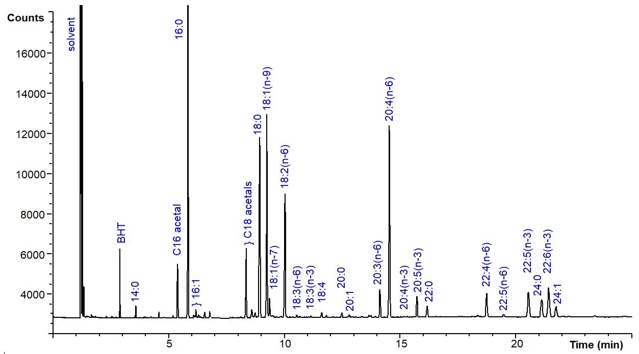
Long chain fatty acids including saturated, monounsaturated, and polyunsaturated species. Quantified by GC-MS with 60-m DB-225 column for isomer resolution. Deuterated IS: d3-C14:0, d3-C16:0, d3-C18:0, d5-C20:4n6.
| Category | Analytes | IS | LLOQ |
|---|
| Saturated LCFA | Myristic acid (C14:0), Pentadecanoic acid (C15:0), Palmitic acid (C16:0), Heptadecanoic acid (C17:0), Stearic acid (C18:0), Arachidic acid (C20:0), Heneicosanoic acid (C21:0) | d3-C14:0 / d3-C16:0 / d3-C18:0 | 0.2–0.5 ng/mL |
| MUFA (ω-7, ω-9) | Palmitoleic acid (C16:1n7), Oleic acid (C18:1n9c), Vaccenic acid (C18:1n7), Eicosenoic acid (C20:1n9) | d3-C16:0 / d3-C18:0 | 0.2–0.5 ng/mL |
| PUFA (ω-6) | Linoleic acid (C18:2n6c), γ-Linolenic acid (C18:3n6), Eicosadienoic acid (C20:2n6), Dihomo-γ-linolenic acid (C20:3n6), Arachidonic acid (C20:4n6), Adrenic acid (C22:4n6) | d5-C20:4n6 | 0.1–0.5 ng/mL |
| PUFA (ω-3) | α-Linolenic acid (C18:3n3), Eicosapentaenoic acid — EPA (C20:5n3), Docosapentaenoic acid — DPA (C22:5n3) | d5-C20:4n6 | 0.1–0.5 ng/mL |
| Trans Fatty Acids | Trans-oleic acid (C18:1t), Trans-linoleic acid (C18:2tt) | d3-C18:0 | 0.3 ng/mL |
GC-MS & LC-MS/MS Method Specifications
| Parameter | Specification |
|---|
| Primary Platform | Agilent 7890A GC with 5977A MSD; 60-m DB-225 column (0.25 mm ID, 0.20 μm film) |
| Derivatization | BF3/MeOH (14% w/v) at 100°C × 30 min; FAME extraction into hexane |
| Confirmatory Platform | SCIEX Triple Quad 6500+ LC-MS/MS with C18 column; direct FFA detection without derivatization |
| Calibration | 7-point matrix-matched with 1/x2 weighted regression; r2 ≥ 0.995 per analyte |
| Precision (CV) | Intra-day <5% (MCFA C8–C12, LCFA C14–C18), <10% (C6:0, PUFA C20+) |
| Recovery | 85–115% at LQC/MQC/HQC for all analytes; IS recovery 80–120% per sample |
| Throughput | 45-min GC temperature program; up to 200 samples per batch |
Extend your analysis with complementary fatty acid panels using matched sample preparation protocols.
Why Choose Our MCFA & LCFA Analysis Platform
- 40+ analytes from C6:0 to C21:0 in a single method — MCFA and LCFA quantified simultaneously with optimized temperature program capturing both volatile and high-boiling FAMEs in one run.
- Deuterated internal standards for absolute quantification — d3-C8:0, d3-C10:0, d3-C12:0 (MCFA) and d3-C14:0, d3-C16:0, d3-C18:0, d5-C20:4n6 (LCFA) spiked pre-extraction.
- 60-m DB-225 column resolves C18:1n9 from C18:1n7 and C18:3n3 from C18:3n6 — isomer-level identification supported by retention index matching against authenticated reference standards.
- Flexible panel selection — MCFA-only (12 analytes), LCFA-only (28+ analytes), or combined MCFA+LCFA panel (40+). Order only what your study needs.
- Dual-platform cross-validation — GC-MS primary quantification with LC-MS/MS confirmation on selected analytes. Concordance data provided for critical studies.
- Split-dilution strategy for wide dynamic range — abundant LCFA (mM) and trace PUFA (sub-μM) quantified from the same sample with cross-dilution verification.
Chain Length Determines Metabolic Fate — Integrating MCFA & LCFA Data
A palmitic acid (C16:0) concentration alone tells you about pool size — not flux, not fate, and not the metabolic context. MCFA and LCFA occupy distinct metabolic compartments: MCFA bypass the carnitine shuttle and drive hepatic ketogenesis; LCFA are incorporated into structural lipids and signaling pathways. Measuring both — and their ratios — reveals which arm of fatty acid metabolism is active in your experimental system.
MCFA — Rapid Oxidation & Ketogenesis
MCFA enter the portal vein directly, reach the liver within minutes, and cross the mitochondrial membrane via simple diffusion — no CPT1 required. This makes them preferential energy substrates that are rapidly oxidized to acetyl-CoA, driving ketone body production when carbohydrate availability is low. MCFA analysis is central to:
- Ketogenic diet mechanism studies (MCT oil → β-hydroxybutyrate)
- Infant formula lipid optimization (MCFA digestibility without pancreatic lipase)
- Sport nutrition (MCFA as rapid energy substrate)
- Gut-liver axis research (MCFA produced by gut microbiota from dietary fiber)
LCFA — Structural Lipids & Signaling
LCFA are esterified into triglycerides and phospholipids, forming the bulk of stored fat and membrane lipid bilayers. Their cellular uptake requires CD36/FATP transporters and CPT1-dependent mitochondrial import — a regulatory step targeted by malonyl-CoA in metabolic disease. LCFA analysis is central to:
- NAFLD/NASH research (palmitate-driven lipotoxicity and ER stress)
- Cardiovascular studies (ω-3/ω-6 balance, arachidonic acid → eicosanoid signaling)
- Membrane lipid remodeling (phospholipid fatty acid composition)
- Inflammation research (arachidonic acid → prostaglandin/leukotriene cascades)
The MCFA/LCFA ratio — particularly C8:0+C10:0 relative to C16:0+C18:0 — provides a functional readout of hepatic fatty acid partitioning. An elevated ratio indicates preferential MCFA oxidation (ketogenic shift); a depressed ratio with elevated C16:0 suggests LCFA-driven lipotoxicity. Neither measurement alone provides this insight — which is why our combined panel includes both.
MCFA & LCFA Analysis Workflow & Instrument Platform

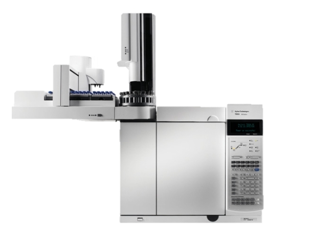
Agilent 7890A GC System (Figure from Agilent)
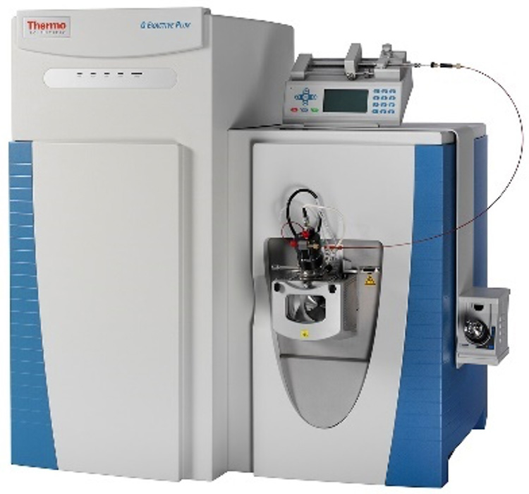
Thermo Fisher Q Exactive (Figure from Thermo Fisher)
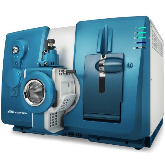
SCIEX Triple Quad 6500+ (Figure from SCIEX)
MCFA & LCFA Analysis — Results and Data Analysis
MCFA & LCFA Quantification Report
Results we provide:
- Absolute concentrations for 40+ fatty acids (μM or μg/mg)
- MCFA/LCFA ratio, ω-3/ω-6 ratio, saturated/unsaturated ratio per sample
- Internal standard recovery and batch QC metrics
- PCA for group clustering and outlier detection
QC metrics included:
- Calibration curve r2, IS recovery, blank carryover per batch
- 3-level QC sample tracking across the analytical sequence
Distribution of detected fatty acid subclasses across experimental groups.
Relative difference in significantly altered fatty acids between groups.
Comparative & Statistical Analysis
Results provided:
- Group comparisons (t-test, ANOVA) with FDR correction per fatty acid
- Volcano plots — fold-change vs significance across all 40+ analytes
- Correlation heatmap across all detected fatty acid species
- ω-3/ω-6 ratio comparison with group-level statistics
Pathway & Multi-Omics Integration
Results provided:
- KEGG fatty acid biosynthesis and β-oxidation pathway mapping
- Integration with fatty acid oxidation and acylcarnitine data
- ML-assisted feature ranking for fatty acid biomarker discovery
- Publication-ready figures (TIFF/PDF, 600 dpi)
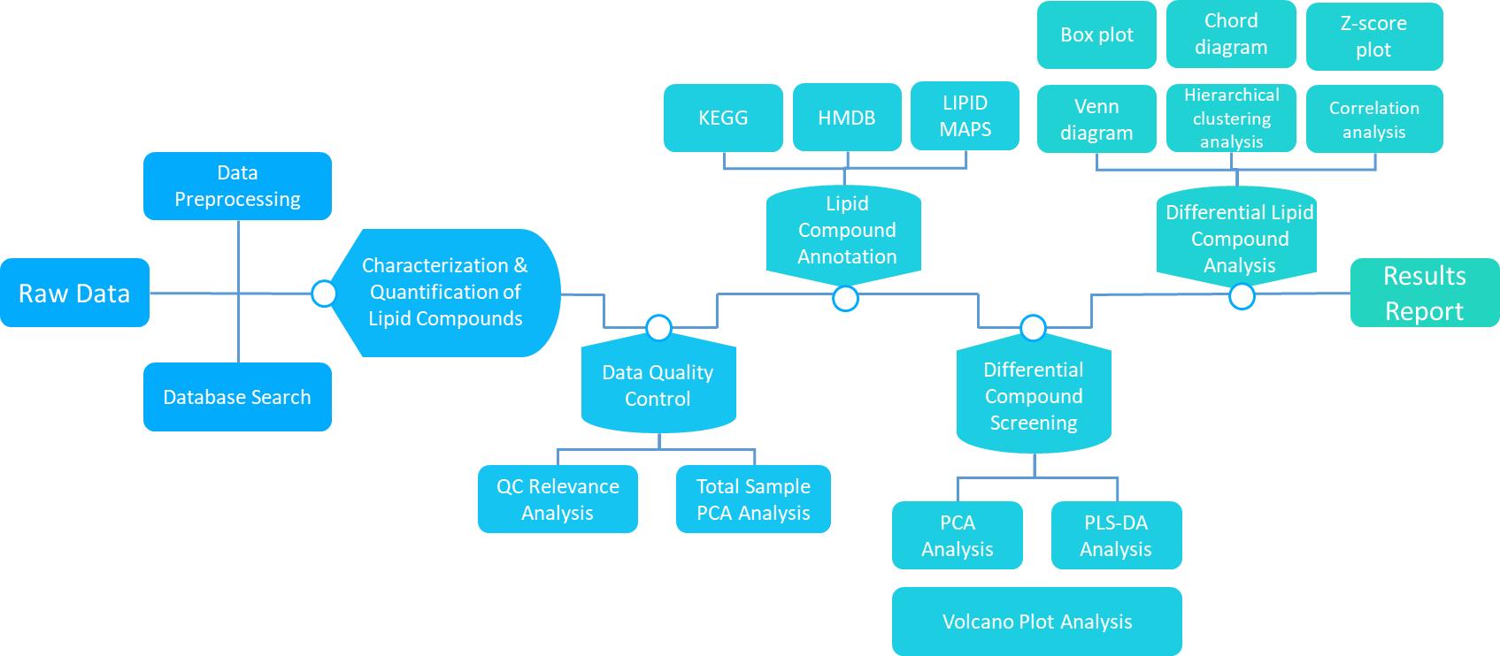 Workflow of Lipidome Data Analysis
Explore our Lipidomics Solutions brochure to learn more about the full spectrum of MCFA, LCFA, and targeted lipidomics analysis capabilities.
Download Brochure
What Our MCFA & LCFA Analysis Used For
Ketogenic Diet & MCT Research
MCFA from MCT oil are rapidly oxidized to ketone bodies. Quantify plasma C8:0, C10:0, and β-hydroxybutyrate in dietary intervention studies, epilepsy models, and neurodegenerative disease research.
NAFLD, Obesity & Metabolic Syndrome
Palmitate (C16:0)-driven lipotoxicity, DAG accumulation, and ER stress are hallmarks of NAFLD/NASH. Tissue and plasma LCFA profiling in dietary and genetic obesity models. Diabetes lipidomics integration available.
Cardiovascular & ω-3/ω-6 Research
EPA (C20:5n3), DHA (C22:6n3), and arachidonic acid (C20:4n6) are precursors to pro- and anti-inflammatory eicosanoids. The ω-3/ω-6 ratio is a key predictor of cardiovascular risk in cohort studies.
Infant Formula & Nutritional Lipidomics
MCFA are critical in infant formulas because neonates have limited pancreatic lipase. Quantify MCFA/LCFA ratios in formula development, breast milk comparison, and weaning studies.
Cancer Metabolism
Tumor cells upregulate de novo fatty acid synthesis — saturated LCFA (C16:0, C18:0) and MUFA (C18:1) are hallmarks of lipogenic tumors. Fatty acid profiling tracks metabolic reprogramming in cancer models. Disease research support available.
Food Quality & Authenticity
Fatty acid profiling for edible oil authentication, trans-fat content verification, and chain-length distribution in functional foods. Our food & nutrition platform supports regulatory-grade documentation.
Sample Requirements for MCFA & LCFA Analysis
| Sample Type | Minimum Amount | Critical Handling |
|---|
| Plasma / Serum | 100 μL | EDTA plasma preferred. Separate within 30 min at 4°C. Snap-freeze in LN2. For MCFA analysis, avoid exposure to room temperature — MCFA are volatile and C6:0–C8:0 can be lost during delayed processing. Store -80°C. |
| Tissue | 50 mg | Snap-freeze in LN2 within 5 min. Perfuse liver with ice-cold saline to remove blood lipids. For adipose tissue, record depot location and freeze immediately to prevent lipase-mediated triglyceride hydrolysis. |
| Cell Pellets | 2 × 10⁶ cells | Wash 2× with ice-cold PBS to remove serum lipids. For MCFA analysis, minimize wash steps — MCFA can leach from cells during PBS incubation. Pellet and snap-freeze. |
| Food / Oil | 50–100 mg | Homogenize under nitrogen. For oils, protect from light and oxygen — PUFA auto-oxidation begins immediately upon air exposure. Add 0.005% BHT to homogenization solvent. |
| Milk / Formula | 100–200 μL | Homogenize before aliquoting — milk fat separates during freezing. For infant formula, record reconstitution method if applicable. Store -80°C. |
FAQ — MCFA & LCFA Analysis
Can I order MCFA-only or LCFA-only instead of the combined panel?
Yes. We offer MCFA-only (C6:0–C12:0, 12 analytes), LCFA-only (C14:0–C21:0, 28+ analytes), and combined MCFA+LCFA (40+ analytes) panels. If your research only involves MCT oil / ketogenic diet endpoints, the MCFA-only panel is sufficient and more cost-effective. For cardiovascular or NAFLD studies focused on long chain fatty acids, the LCFA panel covers saturated, MUFA, PUFA, and trans species.
How do you prevent MCFA loss during sample preparation?
C6:0–C10:0 are volatile — up to 50% can be lost during standard nitrogen blow-down. We use low-temperature (30°C) evaporation with trimethylamine as a co-solvent to reduce vapor pressure. Deuterated MCFA internal standards (d3-C8:0, d3-C10:0, d3-C12:0) are spiked before extraction and undergo the same evaporative conditions, mathematically correcting for any residual loss.
Can you separate oleic acid (C18:1n9) from vaccenic acid (C18:1n7)?
Yes. Our 60-m DB-225 column (bis-cyanopropyl phase, 0.25 mm ID, 0.20 μm film) achieves baseline resolution of C18:1n9c from C18:1n7, C18:2n6c from C18:2tt, and C18:3n3 from C18:3n6. These isomers co-elute on standard 30-m columns and are commonly misreported as a single peak by labs using shorter columns.
What derivatization method do you use — BF3 or HCl/MeOH?
We use BF3/MeOH (14% w/v, 100°C × 30 min) as our primary derivatization method. BF3 provides faster and more complete methylation of both free fatty acids and esterified fatty acids compared to HCl/MeOH, and is the recommended method by AOCS (Ce 2-66). For samples containing cyclopropane or epoxy fatty acids that are sensitive to BF3, we offer HCl/MeOH as an alternative.
Do you report free fatty acids, total fatty acids, or both?
Our standard panel reports total fatty acids (after saponification and methylation of all lipid classes). This gives you the complete fatty acid composition across triglycerides, phospholipids, and cholesterol esters. Free (non-esterified) fatty acid quantification is available as an add-on using a separate extraction without saponification. Contact us to discuss which is appropriate for your study.
How does GC-MS compare to LC-MS/MS for fatty acid analysis?
GC-MS is our primary platform: superior chromatographic resolution for FAME isomers (60-m column), well-established NIST/Wiley spectral libraries for identification, and 45-min temperature program with baseline separation of 40+ FAMEs. LC-MS/MS is our confirmatory platform: direct detection of underivatized fatty acids with no derivatization bias, and MRM sensitivity for low-abundance PUFA. We recommend GC-MS for comprehensive profiling and LC-MS/MS when derivatization is a concern. Dual-platform cross-validation is available for critical studies.
Is this service for clinical diagnostic use?
No. Our MCFA & LCFA analysis is for research use only (RUO) and is not CLIA-certified or CAP-accredited for clinical diagnostic purposes. Our service supports preclinical and translational research — metabolic disease, nutrition, cardiovascular, and cancer studies — not patient care.
What is the turnaround time and minimum sample count?
Standard turnaround is 2–4 weeks. Up to 50 samples: ~2 weeks; 50–200: ~3 weeks; 200+: ~4 weeks. No minimum sample count — pilot batches are welcome. Expedited 1-week service available. Click "Request Analysis" for study design consultation and a formal quotation.