Antimicrobial Resistance Lipidomics

Antimicrobial Resistance and Bacterial Membrane Biology Lipidomics Service

Creative Proteomics provides high-resolution, mass spectrometry-based lipidomics to elucidate the precise molecular mechanisms of antimicrobial resistance (AMR) and bacterial membrane biology. We empower microbiologists, pharmacologists, and infectious disease researchers to transition from basic minimum inhibitory concentration (MIC) assays to deep molecular mechanism mapping, delivering absolute quantification of bacterial envelope remodeling, membrane permeability, and host-pathogen lipid interactions.

Key capabilities

  • Drug-Resistant Strain Lipid Profiling: Compare the baseline lipidomes of susceptible versus multidrug-resistant (MDR) clinical isolates to identify novel lipid resistance biomarkers.
  • Membrane Remodeling & Permeability Analysis: Quantify shifts in bacterial phospholipids and fatty acid saturation that reduce drug influx or enhance efflux pump efficiency.
  • Antimicrobial Mechanism of Action (MoA) Validation: Track dynamic, time-course lipid degradation events triggered by novel antibiotics or membrane-targeting antimicrobial peptides (AMPs).
Request Analysis

Submit Your Request Now

×

Our services have earned the trust of companies, schools, and organizations globally, and we remain dedicated to maintaining that trust.

Boston University
University at Buffalo
UMass Medical School
Hawaii Pacific University
Medizinische Fakultät
Louisiana State University
Nyulangone
Nature's Fynd
Dietary Supplement Manufacturer
  • Trends & Challenges
  • Lipidomic Workflows
  • Technical Advantages
  • Preclinical Screening
  • Case Studies
  • FAQ

Lipidomic Workflows for Antimicrobial Resistance Research

Deciphering the molecular basis of AMR requires moving beyond simple genomic screening. Select your specific experimental scenario below.

Susceptible vs. Resistant Strain Comparison

Situation

Screening paired clinical isolates (e.g., wild-type vs. MDR P. aeruginosa) to identify the baseline phenotypic mechanisms driving overall drug resistance.

Goal

Conduct an unbiased comparison of the global lipidome to lock in on the specific structural lipid classes uniquely upregulated in the resistant strain.

Recommended path

Discovery → Validation

Recommended services
What you will get

A comprehensive metabolic fingerprint that identifies completely novel lipid biomarkers of resistance, highlighting alternative survival pathways missed by transcriptomics.

Mechanism of Action (MoA) for Novel Antimicrobials

Situation

Evaluating the exact bactericidal mechanism of a newly synthesized antimicrobial peptide (AMP) or small molecule designed to target the bacterial envelope.

Goal

Validate how the drug physically binds to and degrades specific target lipids, leading to fatal membrane depolarization and cell lysis.

Recommended path

Targeted Validation

Recommended services
What you will get

Time-course quantitative data proving the specific, progressive destruction of the bacterial phospholipid bilayer triggered by the drug candidate.

Colistin and Polymyxin Resistance Mechanisms

Situation

Investigating Gram-negative pathogens expressing the mcr-1 gene, which confers resistance to last-resort polymyxin antibiotics.

Goal

Absolutely quantify the specific enzymatic modifications of Lipid A (e.g., addition of phosphoethanolamine) that reduce the negative charge of the outer membrane.

Recommended path

Discovery → Validation

Recommended services
What you will get

High-resolution mass spectra definitively proving the structural alteration of the lipopolysaccharide (LPS) anchor, confirming the functional activity of the resistance gene.

Membrane Permeability and Efflux Pump Function

Situation

Evaluating how bacteria dynamically alter their membrane fluidity to physically restrict antibiotic entry or enhance the operational efficiency of multi-drug efflux pumps.

Goal

Analyze the exact ratio of saturated to unsaturated fatty acids and the remodeling of bulk phospholipids under continuous drug pressure.

Recommended path

Targeted Validation

Recommended services
What you will get

Concrete mathematical indices of membrane saturation and fluidity, directly correlating the biophysical state of the lipid bilayer with reduced intracellular drug concentrations.

Host-Pathogen Interaction and Intracellular Survival

Situation

Studying how intracellular pathogens (like M. tuberculosis or Salmonella) hijack the host's lipid metabolism to survive inside macrophage phagosomes.

Goal

Track the exchange and utilization of specific host lipids (such as cholesterol or sphingolipids) by the invading bacteria to build protective vacuoles.

Recommended path

Discovery → Deep Insight

Recommended services
What you will get

A mapped network distinguishing the bacterial lipidome from the host lipidome, revealing the specific metabolic theft required for pathogen persistence.

Persister Cell and Biofilm Lipid Alterations

Situation

Analyzing chronic infections driven by bacterial biofilms or dormant persister cells that are physically impenetrable by standard antibiotic therapies.

Goal

Define the unique, low-metabolism lipid signature of the biofilm matrix and compare it against the lipidome of actively dividing planktonic cells.

Recommended path

Discovery → Validation

Recommended services
What you will get

Identification of the specific structural lipids required to maintain biofilm architecture, providing novel targets for biofilm-dispersing adjuvant therapies.

Bacterial Membrane Remodeling Case Studies

Reference: Heme alters biofilm formation in Mycobacterium abscessus. Microbiology Spectrum, 2024. DOI: 10.1128/spectrum.02415-24

Heme-Associated Biofilm Remodeling in Mycobacterium abscessus

Project Focus

This client publication is relevant to teams studying how host-associated factors reshape bacterial physiology during chronic infection. In this study, extracellular heme altered the growth pattern and biofilm behavior of Mycobacterium abscessus, a pathogen closely associated with persistent infection and reduced antibiotic susceptibility.

What Was Measured

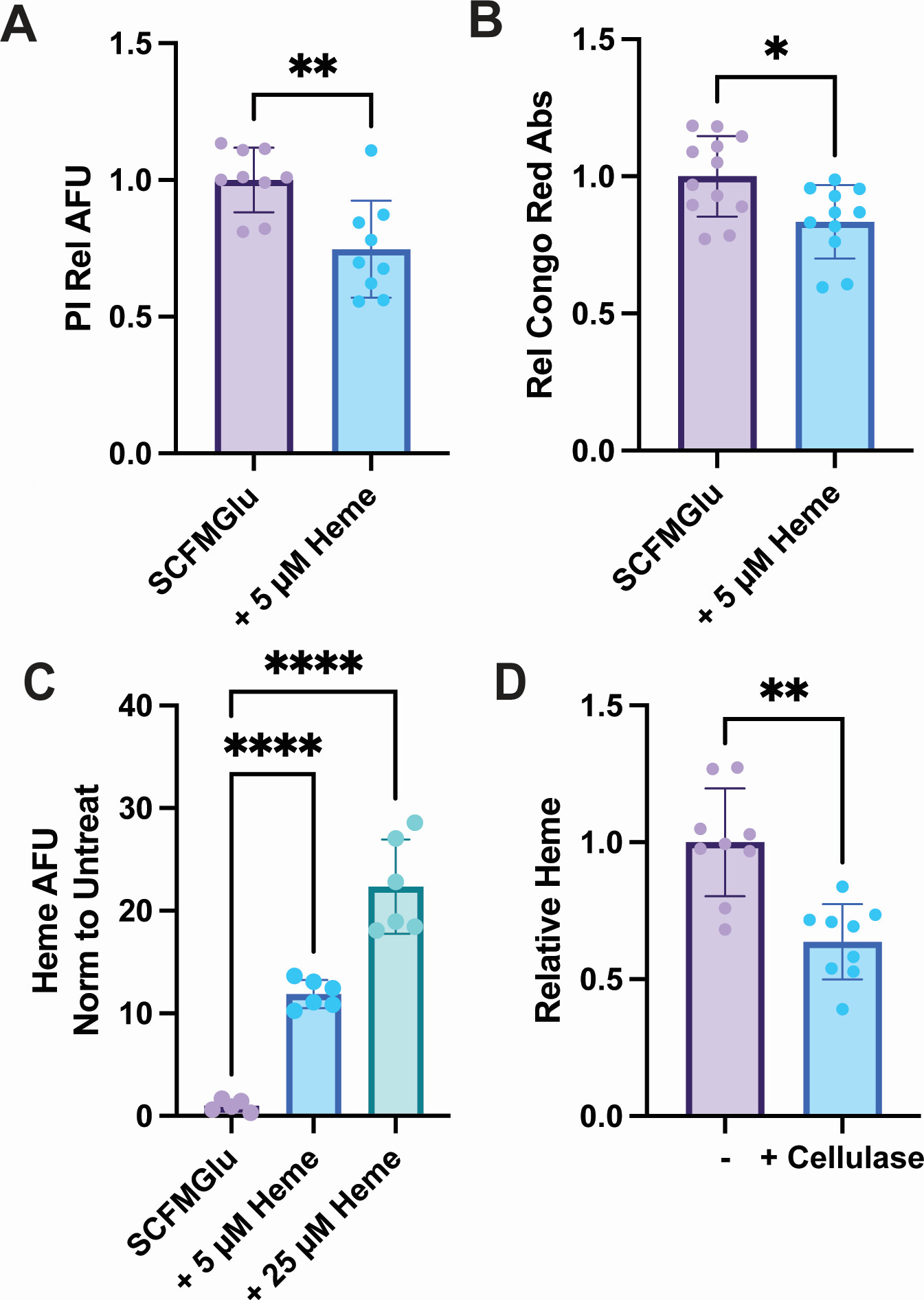
The study examined how exogenous heme influenced growth behavior, biofilm formation, and broader molecular responses in M. abscessus. The published work reports proteomics and metabolomics evidence, making this a strong case for biofilm- and membrane-associated molecular profiling, rather than a strictly lipidomics-only project.

What the Study Showed

The authors found that extracellular heme changed biofilm formation and broader metabolic behavior in M. abscessus. For service-page readers, the value of this case is that it shows how molecular profiling can help explain bacterial adaptation in persistent, barrier-associated states that are difficult to characterize with phenotype assays alone.

Why It Matters
  • Relevant to biofilm-associated infection research
  • Useful for studies of persistence and reduced drug susceptibility
  • Supports omics-guided interpretation of bacterial adaptation under host-like stress
Biofilm assay image from a client publication showing heme-associated growth and biofilm remodeling in Mycobacterium abscessus.
Biofilm phenotype readout showing how extracellular heme changes growth behavior in Mycobacterium abscessus.
Reference: Colistin Treatment Affects Lipid Composition of Acinetobacter baumannii. Antibiotics, 2021. DOI: 10.3390/antibiotics10050528

Lipid Composition Shifts Under Colistin Stress in Acinetobacter baumannii

Project Focus

This publication is highly relevant to antimicrobial resistance and membrane biology research because it examines how a clinically important Gram-negative pathogen changes lipid composition under colistin exposure.

What Was Measured

The study used LC-HRMS²-based untargeted lipidomics to compare susceptible and multidrug-resistant A. baumannii strains under lethal and sublethal colistin treatment. The authors profiled glycerolipids and related membrane-associated lipid changes linked to antibiotic stress adaptation.

What the Study Showed

The paper reports that phosphatidylethanolamines were significantly reduced under treatment, while abundant PE 34:1 and PG 34:1 species increased. For a service page, this is a strong proof point that lipidomics can reveal measurable membrane remodeling under polymyxin pressure and add mechanistic clarity beyond susceptibility testing alone.

Why It Matters
  • Relevant to colistin and polymyxin resistance studies
  • Useful for membrane-active antimicrobial mechanism research
  • Supports lipid-level interpretation of susceptible versus resistant adaptation states
PCA plot from a published lipidomics study showing membrane lipid differences in susceptible and resistant Acinetobacter baumannii strains.
PCA of the bacterial lipidome showing separation between susceptible and resistant A. baumannii under colistin treatment.

Frequently Asked Questions (FAQ)

How does lipidomics reveal novel antibiotic resistance mechanisms missed by genomics?
Genomics tells you what a bacterium is computationally capable of doing; lipidomics tells you what it is actually doing to survive. Many AMR phenotypes arise not from direct target mutations, but from biophysical phenotypic adaptations—like radically shifting the ratio of saturated to unsaturated fatty acids in the membrane to physically block drug entry. Only direct lipidomic measurement can capture these physical barrier defenses.
Can you detect specific lipid A modifications associated with colistin resistance?
Yes. Resistance to polymyxins and colistin frequently involves the enzymatic addition of phosphoethanolamine (PEtN) or 4-amino-4-deoxy-L-arabinose (L-Ara4N) to Lipid A, reducing its net negative charge. Using high-resolution mass spectrometry and targeted fragmentation patterns, we can confidently identify and absolutely quantify these specific, resistance-conferring structural modifications on the intact molecule.
What is the minimum bacterial biomass (e.g., CFU or OD600) required for comprehensive lipid profiling?
Thanks to the extreme sensitivity of our LC-MS/MS platforms (achieving pg/mL LLOQ), we require very low sample inputs. Typically, a washed bacterial pellet derived from 2 to 5 mL of liquid culture at an OD600 of 1.0 (approximately 10^8 to 10^9 CFU) provides more than enough biomass for deep, absolute quantitative lipid profiling of both the inner and outer membranes.
How do you resolve complex bacterial phospholipids like cardiolipin and phosphatidylglycerol (PG)?
Bacterial cardiolipins are massive, tetra-acylated lipids that form incredibly complex mixtures of positional isomers. We utilize optimized, ultra-long reverse-phase UHPLC gradients specifically tailored for large, hydrophobic molecules, coupled with highly specific Multiple Reaction Monitoring (MRM) transitions. This achieves baseline chromatographic separation of critical isomers that standard assays cannot distinguish.
Can your platform differentiate between the lipidomes of persister cells, biofilms, and planktonic bacteria?
Absolutely. We employ customized extraction protocols designed to gently strip away the extracellular polymeric substance (EPS) of biofilms, allowing us to isolate the intact persister cells within. By comparing these cells against free-floating planktonic controls, we can precisely map the metabolic downregulation and specific membrane fortifications unique to the dormant, highly tolerant persister state.
How do you prevent ex vivo lipid degradation when harvesting drug-treated bacterial cultures?
When harvesting bacteria exposed to sub-lethal antibiotic stress, endogenous phospholipases act aggressively. We enforce strict protocols requiring rapid centrifugation followed instantly by sub-second flash freezing in liquid nitrogen. We then utilize chilled, biphasic extraction solvents pre-loaded with antioxidant shields, instantly denaturing all degradative enzymes and freezing the true in vivo stress phenotype.
Can you integrate bacterial lipidomics data with transcriptomics for multi-omics MoA studies?
Yes. To fully elucidate an antimicrobial's Mechanism of Action (MoA), it is crucial to link initial gene expression changes to the final physical destruction of the membrane. Through advanced bioinformatics and network topology modeling, we mathematically integrate your transcriptomic (RNA-Seq) data with our quantitative lipidomic shifts, establishing a clear, multi-tiered pathway of drug-induced bacterial death.
Do you support the analysis of clinical isolates and multidrug-resistant (MDR) pathogens?
Yes. We routinely process and analyze complex clinical isolates, including the challenging ESKAPE pathogens. As previously mentioned, our extraction workflows are robustly designed to process completely inactivated samples, handling organisms with thick, highly modified capsules typical of dangerous MDR clinical strains, ensuring precise data recovery in biosafety-compliant environments for research use.
* Our services can only be used for research purposes and Not for clinical use.

Applications:


Online Inquiry

CONTACT US

Copyright © 2026 Creative Proteomics. All rights reserved.