Neuro-Metabolism Lipidomics for Brain Health Research

Neurodegeneration research increasingly points to lipid remodeling as a measurable layer of neuro-metabolism—both systemically (blood) and locally (brain tissue). Our neurodegeneration lipidomics service supports plasma lipidomics biomarker discovery and brain tissue lipidomics analysis across AD and PD programs, from broad discovery to quantitative validation and spatial context.

Key capabilities

  • End-to-end plasma lipidomics biomarker discovery: untargeted profiling → targeted confirmation → panel-ready outputs
  • Mechanism-focused modules: sphingolipidomics LC-MS/MS and oxysterol profiling LC-MS/MS for pathway interrogation
  • Tissue context: MALDI imaging lipidomics to map lipid signatures to brain regions and ROIs
Request Analysis

Submit Your Request Now

×

Our services have earned the trust of companies, schools, and organizations globally, and we remain dedicated to maintaining that trust.

Boston University
University at Buffalo
UMass Medical School
Hawaii Pacific University
Medizinische Fakultät
Louisiana State University
Nyulangone
Nature's Fynd
Dietary Supplement Manufacturer
  • Trends & Challenges
  • Integrated Solutions
  • Technical Advantages
  • Case Studies
  • FAQ

Solution Matrix For Situation-Triggered Decisions

Neuro-metabolism projects in Alzheimer's disease (AD) and Parkinson's disease (PD) research typically progress through structured analytical stages—from broad lipidome profiling to quantitative validation and, when appropriate, spatial or pathway-focused expansion.

The framework below outlines common study scenarios and aligns them with corresponding workflows across Discovery (Bundle A), Validation (Bundle B), and Deep Insight (Bundle C). This structured approach supports plasma and brain tissue lipidomics studies at different stages of translational research, while maintaining methodological consistency and data interpretability.

Signals Are Weak In Plasma

Situation

Untargeted plasma screening identifies limited statistically robust features, with high inter-individual variability and unclear lipid-class drivers.

Goal

Increase coverage and confidence in candidate selection prior to committing to targeted validation.

Recommended Path

Discovery (Bundle A) → Expanded statistical and pathway-level refinement

Recommended Services

What You Will Receive

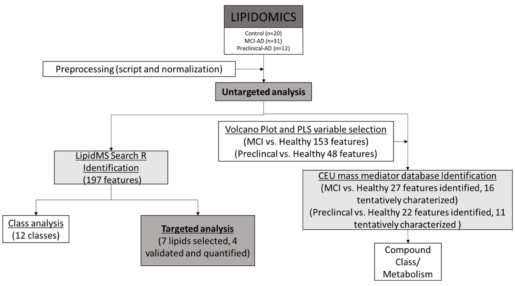
A QC-reviewed differential feature table, multivariate plots (PCA/heatmap), class-level enrichment summaries, and a prioritized lipid shortlist suitable for follow-up validation.

Cross-Batch Replication Is Inconsistent

Situation

Candidate lipids identified during discovery do not reproduce across analytical batches, collection waves, or collaborating sites.

Goal

Stabilize quantification and establish a reproducible lipid panel for cohort-level comparison.

Recommended Path

Validation (Bundle B) → Batch-aware statistical reporting

Recommended Services

What You Will Receive

Quantitative LC-MS/MS outputs with internal standard QC review, cross-batch comparability assessment, and a refined marker panel with improved analytical stability.

Sphingolipid Pathways Require Quantitative Confirmation

Situation

Genetic, transcriptomic, or prior literature evidence implicates sphingolipid or ceramide metabolism in AD/PD biology.

Goal

Quantify species-level sphingolipid changes to support mechanistic interpretation.

Recommended Path

Discovery screening → Targeted confirmation using focused sphingolipid panels

Recommended Services

What You Will Receive

Quantitative sphingolipid and ceramide datasets, species-level concentration tables, and pathway-aligned summaries linking plasma and brain tissue findings.

Spatial Localization Is Required In Brain Tissue

Situation

Bulk lipidomics indicates significant changes, but regional specificity within brain tissue remains unresolved.

Goal

Determine anatomical distribution of lipid alterations and correlate with defined regions or histological features.

Recommended Path

Targeted validation → Spatial lipid mapping (Deep Insight)

Recommended Services

What You Will Receive

Spatial ion intensity maps, ROI-level comparisons, and region-resolved lipid distribution profiles suitable for integration with histological or molecular datasets.

Cholesterol Turnover And Oxysterols Are Implicated

Situation

Data or literature suggests altered cholesterol metabolism, oxidative stress, or sterol oxidation in AD/PD models.

Goal

Quantify oxysterols and evaluate cholesterol pathway remodeling within plasma or brain tissue.

Recommended Path

Targeted sterol module → Optional spatial confirmation if tissue context is required

Recommended Services

What You Will Receive

Oxysterol quantification tables, cholesterol pathway summaries, and analytically validated sterol profiles supporting neuro-metabolic interpretation.

Mitochondrial Lipid Remodeling Is Suspected

Situation

Preliminary data indicate bioenergetic stress or membrane remodeling consistent with mitochondrial involvement.

Goal

Differentiate systemic lipid alterations from mitochondria-associated remodeling and prioritize confirmatory markers.

Recommended Path

Discovery profiling → Mitochondria-focused analysis → Targeted confirmation

Recommended Services

What You Will Receive

Mitochondria-enriched lipid profiles, mechanistic pathway interpretation, and a focused marker set for downstream validation.

Limited Brain Tissue Input

Situation

Microdissected regions or archival samples restrict available tissue mass.

Goal

Maximize analytical yield while maintaining data robustness and interpretability.

Recommended Path

Fit-for-sample discovery workflow → Focused targeted validation

Recommended Services

What You Will Receive

An optimized analytical plan aligned to input constraints, supported by targeted confirmation to reduce false positives and enhance interpretability.

Directionality Of Lipid Changes Must Be Clarified

Situation

Abundance data alone cannot determine whether lipid changes reflect altered synthesis, turnover, or accumulation.

Goal

Introduce metabolic context to support mechanistic conclusions.

Recommended Path

Metabolic flux assessment → Targeted confirmation of key markers

Recommended Services

What You Will Receive

Turnover-oriented analytical outputs that clarify pathway directionality and support mechanistic interpretation in AD/PD neuro-metabolism studies.

Case Studies

Publication: Peña-Bautista et al., 2022, Journal of Clinical Medicine. https://doi.org/10.3390/jcm11175030
Who needs this: Translational AD teams comparing controls vs preclinical/MCI due to AD using blood-based readouts.

Case 1 — Plasma Lipidomics For Early-Stage AD Stratification

Method used

Untargeted LC-MS lipidomics on plasma across clinical groups, followed by targeted analysis for selected lipids.

Result obtained

Reports statistically significant differences across lipid families (e.g., DG, LPE, LPC, MG, SM) and highlights a candidate lipid species for follow-up validation.

Recommended path

A → B

Workflow schematic for plasma lipidomics biomarker discovery in early Alzheimer's disease research (LC-MS/MS).
Study workflow from untargeted plasma lipidomics biomarker discovery to targeted confirmation in early AD research.
Publication: Krokidis et al., 2024, Cells. https://doi.org/10.3390/cells13080702
Who needs this: Teams exploring whether extracellular vesicles offer a brain-informative lipid layer beyond bulk plasma.

Case 2 — Plasma EV Lipidomics To Probe Brain-Linked Lipid Signatures In AD (RUO)

Method used

Targeted lipidomic analysis in plasma-derived EV subpopulations (including neuronal- and astrocytic-enriched captures) comparing AD vs controls after lipoprotein removal.

Result obtained

Reports lipid-class and species-level differences across EV subtypes, supporting EV lipidomics as a mechanism-oriented biomarker discovery approach.

Recommended path

B → C (if spatial/tissue mapping is needed

Percent-difference plots of EV lipid classes in Alzheimer's disease vs control using targeted lipidomics LC-MS/MS.
Lipid class shifts across plasma EV subtypes comparing Alzheimer's disease and control samples in targeted lipidomics.
Publication: Cheng et al., 2011, PLOS ONE. https://doi.org/10.1371/journal.pone.0017299
Who needs this: Neurodegeneration teams seeking brain tissue lipidomics analysis that links lipid pathways with region-specific PD biology.

Case 3 — Brain Tissue Lipid Pathway Remodeling In PD Visual Cortex

Method used

LC-MS lipidomics screening across ~200 lipid species in human brain regions (PD vs controls), plus targeted oxysterol measurements and gene expression context.

Result obtained

Reports extensive lipid changes in visual cortex (including sphingolipid/glycerophospholipid and oxysterol alterations) and provides heatmap-style summaries for pathway interpretation.

Recommended path

A → B → C

Heatmap summarizing differential lipid species in Parkinson's disease brain tissue lipidomics analysis (LC-MS).
Heatmap of PD-associated lipid changes detected by LC-MS lipidomics in human brain tissue comparisons.

FAQ

How can I minimize artifactual lipid oxidation in polyunsaturated-rich brain samples?
Brain tissue is a high-performance lipid organ, making it exceptionally prone to oxidation. To ensure biological accuracy, we implement a cold-chain extraction protocol including rapid liquid nitrogen freezing and processing at sub-zero temperatures. The addition of antioxidants like BHT during organic-phase extraction and the use of amber vials for light-sensitive oxysterols are mandatory. These steps ensure that detected oxidative markers, such as specific sterols or PUFA derivatives, reflect actual neuropathology rather than benchtop degradation.
Why do lipidomic signatures often fail to replicate across different clinical plasma cohorts?
High inter-individual variability and the blood–brain barrier often dilute CNS-specific signals in bulk plasma. Inconsistency is typically mitigated by shifting from relative abundance measurements to internal-standard-based absolute quantification. For deeper insight, we recommend isolating neuron-derived extracellular vesicles to capture brain-specific lipid cargoes. This approach bypasses systemic metabolic noise and significantly improves the reproducibility of markers intended to reflect central nervous system health.
How can I distinguish between structurally similar sphingolipid isomers in AD research?
Many sphingolipid subclasses share near-identical masses, which can lead to misidentification in broad screenings. Our workflow utilizes LC–MS/MS with specific diagnostic fragment monitoring and retention-time indexing. By targeting unique fragments, such as m/z 264.3 for sphingosine-based species, we eliminate isobaric interference. This precision allows researchers to map changes to specific enzymatic pathways, such as the de novo synthesis pathway, with high confidence in the identity of each lipid species.
When is it necessary to move from bulk brain lipidomics to MALDI imaging?
Transition to MALDI-MSI when regional specificity is required to address the spatial dimension of a lipid shift. While bulk lipidomics provides deep coverage, it averages signals across distinct anatomical zones. If your hypothesis involves lipid remodeling specifically within hippocampal subfields or around amyloid plaques, spatial mapping at 5–20 μm resolution is necessary. This enables direct correlation of lipid alterations with localized neuroinflammation or histological hallmarks.
Can meaningful data be extracted from microdissected brain regions under 5 mg?
Yes. Limited input requires a high-sensitivity targeted workflow rather than broad discovery screening. By optimizing ion-source parameters and utilizing micro-flow LC–MS/MS, we reach picomolar detection limits. For archival or microdissected samples, we prioritize the most biologically relevant classes to maximize data density from minimal biomass. This ensures that low-abundance regulatory lipids, such as signaling phospholipids, are quantified without compromising data robustness.
Does an increase in lipid abundance always indicate upregulated synthesis in neurodegeneration?
Static concentration data cannot distinguish between lipid accumulation due to impaired clearance and increased synthesis. To resolve directionality, we employ stable isotope-labeled (13C or D₂O) metabolic flux analysis. By tracking the incorporation of labeled precursors into lipid species, we calculate the fractional synthesis rate. This provides the necessary metabolic context to determine whether a pathway is truly overactive or whether the brain is failing to clear metabolic byproducts.
* Our services can only be used for research purposes and Not for clinical use.

Applications:


Online Inquiry

CONTACT US

Copyright © 2026 Creative Proteomics. All rights reserved.