Our services have earned the trust of companies, schools, and organizations globally, and
we remain dedicated to maintaining that trust.
What Is Exosome Lipidomics?
Exosomes and extracellular vesicles (30–150 nm) are released by virtually all cell types. Their lipid bilayer
reflects the endosomal sorting processes that generated the vesicle, encodes information about the parent
cell's metabolic state, and mediates interactions with recipient cells through lipid receptors and membrane
fusion events.
Exosome lipidomics profiles the lipid complement of purified EVs — both by untargeted LC-MS/MS for discovery
(300+ species without pre-selection) and by targeted MRM for absolute quantification of specific lipid panels. The
single largest source of error in either approach is not the mass spectrometer: it is the EV isolation method,
which co-purifies non-exosomal lipids indistinguishable from genuine EV signals.
How EV Isolation Method Affects Lipidomics Data
— and Our Controls
Each isolation technique carries a characteristic contaminant profile. Understanding these is essential for
distinguishing EV biology from isolation artifact.
| Isolation Method |
Co-Isolated Lipid Contaminants |
Mitigation Strategy |
| Ultracentrifugation (UC) |
LDL, VLDL, chylomicron remnants co-pelleted at 100,000×g — falsely elevates cholesterol esters and
triglycerides |
Iodixanol density gradient after UC pelleting; buoyant density verification |
| Size Exclusion Chromatography (SEC) |
HDL, free cholesterol, albumin-bound fatty acids co-eluting in early EV fractions |
Fraction-level profiling — compare EV-rich vs soluble SEC fractions to identify EV-enriched lipids |
| Polymer Precipitation (PEG) |
Non-EV proteins, aggregated lipoproteins, lipid micelles — up to 80% of lipid signal from non-EV sources
|
Not recommended for lipidomics. If PEG isolation is unavoidable, SEC polishing is required |
| Immunoaffinity Capture |
Elution buffer detergents, antibody-derived contaminants |
Detergent-free elution; blank capture control run in parallel for background subtraction |
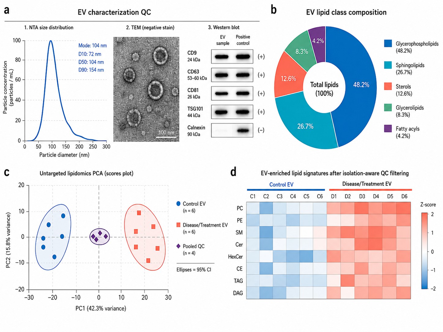
Every project undergoes EV characterization before lipid extraction: nanoparticle tracking analysis (NTA) for
size and concentration, transmission electron microscopy (TEM) for morphology, and Western blot for EV markers
(CD9, CD63, CD81, TSG101) and purity markers (calnexin, GM130). These QC data are included in your report.
Exosome Lipidomics Service in Creative Proteomics
We offer both untargeted discovery and targeted validation lipidomics for purified exosome and EV samples,
supporting all major isolation methods with EV characterization QC included. Choose the approach that matches your
research stage, or combine both in a discovery-to-validation workflow.
Untargeted Discovery Lipidomics
High-resolution LC-MS/MS (Thermo Q Exactive HF-X) with data-dependent acquisition in both positive and
negative ion modes. LipidSearch identification against LIPID MAPS and HMDB. Profiles 300+ lipid species
across glycerophospholipids, sphingolipids, sterols, glycerolipids, and fatty acyls from a single
injection.
Targeted MRM Quantification Panels
Absolute quantification of user-selected lipid classes using SCIEX Triple Quad 6500+ with scheduled MRM
and stable isotope internal standards. Ceramides (C16:0–C24:1), sphingomyelins, phospholipids (PC, PE,
PS, PI), cholesterol esters, and free fatty acids quantified at sub-picomole LLOQ with CV below 10%.
EV Characterization Before Lipid Extraction
NTA for size and concentration, TEM for morphology verification, and Western blot for EV markers (CD9,
CD63, CD81, TSG101) and purity (calnexin). Performed before lipid extraction on every sample —
characterization data are included in the final report.
Isolation-Method-Aware Processing
Your EV isolation method is factored into data analysis. Fraction-level comparison (for SEC) and
density gradient verification (for UC) distinguish genuine EV lipids from co-isolated contaminants.
Method-specific background controls included in every batch.
Multi-Omics Integration
EV lipidomics data formatted for integration with proteomics and transcriptomics. Correlation networks
linking EV lipid species to EV protein cargo and parent-cell gene expression. Compatible with pathway
analysis in KEGG, Reactome, and WikiPathways.
EV Lipid Classes & Targeted Panel Options
- Untargeted Coverage
- Targeted Panels
- Method Specs
Untargeted Profiling — Lipid Classes Detected
Data-dependent acquisition in positive and negative ion modes. LipidSearch identification with manual
spectral verification. Typical coverage: 300+ lipid species.
| Lipid Class |
Subclasses Detected |
Typical Species |
| Glycerophospholipids |
PC, PE, PS, PI, PG, PA, CL, LPC, LPE, ether-linked (PC O-, PE O-) |
120+ |
| Sphingolipids |
Cer, SM, HexCer, LacCer, GM3, Sulf, dhCer, CerP, S1P |
60+ |
| Sterol Lipids |
Cholesterol, cholesteryl esters, oxysterols |
15+ |
| Glycerolipids |
TAG, DAG, MAG |
80+ |
| Fatty Acyls |
Free fatty acids, acyl-carnitines, eicosanoids |
30+ |
Targeted MRM Quantification Panels
Absolute quantification with stable isotope-labeled internal standards on SCIEX Triple Quad 6500+. Select
individual panels or combine for broader coverage. Each panel reports absolute concentrations (nmol/mL or
pmol/μg protein) with CV below 10%.
| Panel |
Analytes |
LLOQ |
| Ceramide Panel |
Cer C16:0, C18:0, C18:1, C20:0, C22:0, C24:0, C24:1; dhCer C18:0, C24:0; Cer1P C16:0, C18:0 |
0.1 pmol/mL |
| Phospholipid Panel |
PC 32:0–40:6, PE 34:1–40:6, PS 36:1–40:6, PI 36:2–38:4, LPC 16:0–18:1, LPE 16:0–18:1 |
0.5 pmol/mL |
| Sphingomyelin Panel |
SM d18:1/16:0, 18:0, 18:1, 20:0, 22:0, 24:0, 24:1 |
0.2 pmol/mL |
| Sterol & Oxysterol Panel |
Cholesterol, CE 18:1, CE 20:4; 7-KC, 7β-HC, 27-HC, 24S-HC |
1.0 pmol/mL |
Method Specifications
| Parameter |
Untargeted |
Targeted |
| Platform |
Thermo Q Exactive HF-X Orbitrap |
SCIEX Triple Quad 6500+ |
| Acquisition |
DDA; m/z 200–2000; resolution 120K (MS1), 30K (MS2) |
Scheduled MRM; 2-3 transitions per analyte |
| Chromatography |
C18 column (2.1 × 100 mm, 1.7 μm); 25-min ACN/IPA/H2O gradient |
| Lipid Extraction |
MTBE-based; SPLASH Lipidomix (untargeted) or analyte-matched deuterated IS (targeted)
|
| Identification |
LipidSearch 5.0; LIPID MAPS + HMDB; manual spectral review |
Retention time + ion ratio matching against authentic standards |
| QC |
Pooled QC every 8 injections; CV below 30% (untargeted) / below 10% (targeted) |
Why Choose Our Exosome Lipidomics Platform
- EV characterization before lipid extraction — NTA, TEM, and Western blot data included in every report,
so you know exactly what particle population was profiled.
- Isolation-method-aware processing — UC, SEC, or immunoaffinity protocols are factored into data analysis
with method-specific background controls.
- Untargeted-to-targeted continuity — discovery hits from untargeted profiling can be directly
transitioned to targeted MRM validation using the same sample preparation workflow.
- LipidSearch identification with manual spectral verification — automated database matching at ppm below
5 followed by expert review to remove false positives.
- Pooled QC every 8 injections with CV filtering — differential signals reflect biology, not analytical
drift.
Data Normalization in EV Lipidomics — Why Reporting Units Matter
A given lipid species can appear upregulated or downregulated depending solely on how you normalize the data.
In EV lipidomics, particle concentration, protein mass, and total lipid content are not proportional across
preparations — especially when comparing samples isolated by different methods or from different biofluids.
Per particle (NTA-normalized)
Lipid abundance expressed per EV particle (mol/particle). Best for comparing EV populations with different
sizes or when particle count is directly measured. Sensitive to NTA precision — variations in particle
counting propagate into lipid ratios. Requires NTA data for each sample.
Per protein (BCA-normalized)
Lipid abundance per μg EV protein. Most commonly used in EV literature and compatible with proteomics
co-analysis. Different EV populations carry different protein-to-lipid ratios; normalization to protein can
mask or exaggerate lipid changes when comparing EVs from different sources.
We report lipid abundance under both normalization strategies as standard, along with
total lipid class composition (mol% per class). For studies comparing EVs across isolation methods or biofluids,
we recommend reviewing data under both strategies before drawing conclusions. This is consistent with ISEV
MISEV2018 guidelines for transparent reporting of normalization methods.
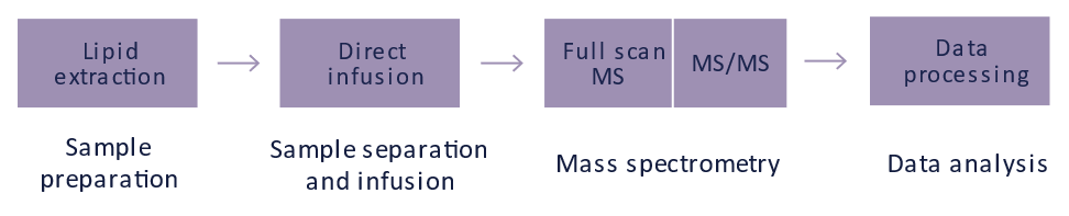
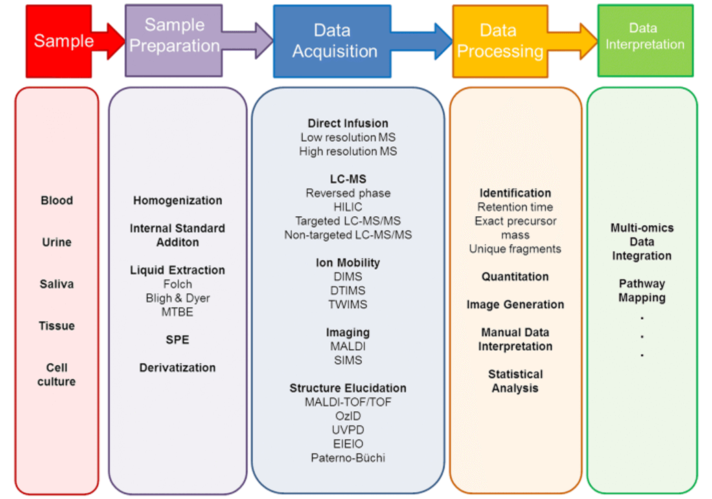
Exosome Lipidomics Workflow
& Instrument Platform
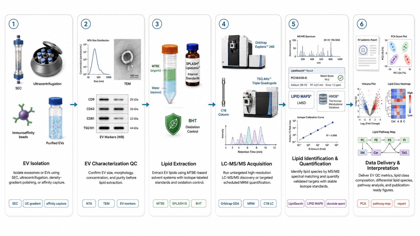
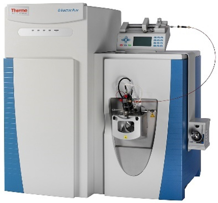
Thermo Q Exactive HF-X Orbitrap (Figure from Thermo Fisher)
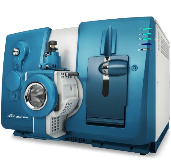
SCIEX Triple Quad 6500+ (Figure from SCIEX)
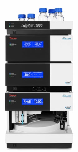
Thermo Scientific UltiMate 3000 (Figure from Thermo Fisher)
Exosome Lipidomics — Results & Data
Analysis
Untargeted Discovery Report
Results provided:
- 300+ identified lipid species with LipidSearch confidence grades
- Retention time, m/z, adduct type, and MS/MS fragment matching
- Relative abundance per lipid species (IS-normalized peak area)
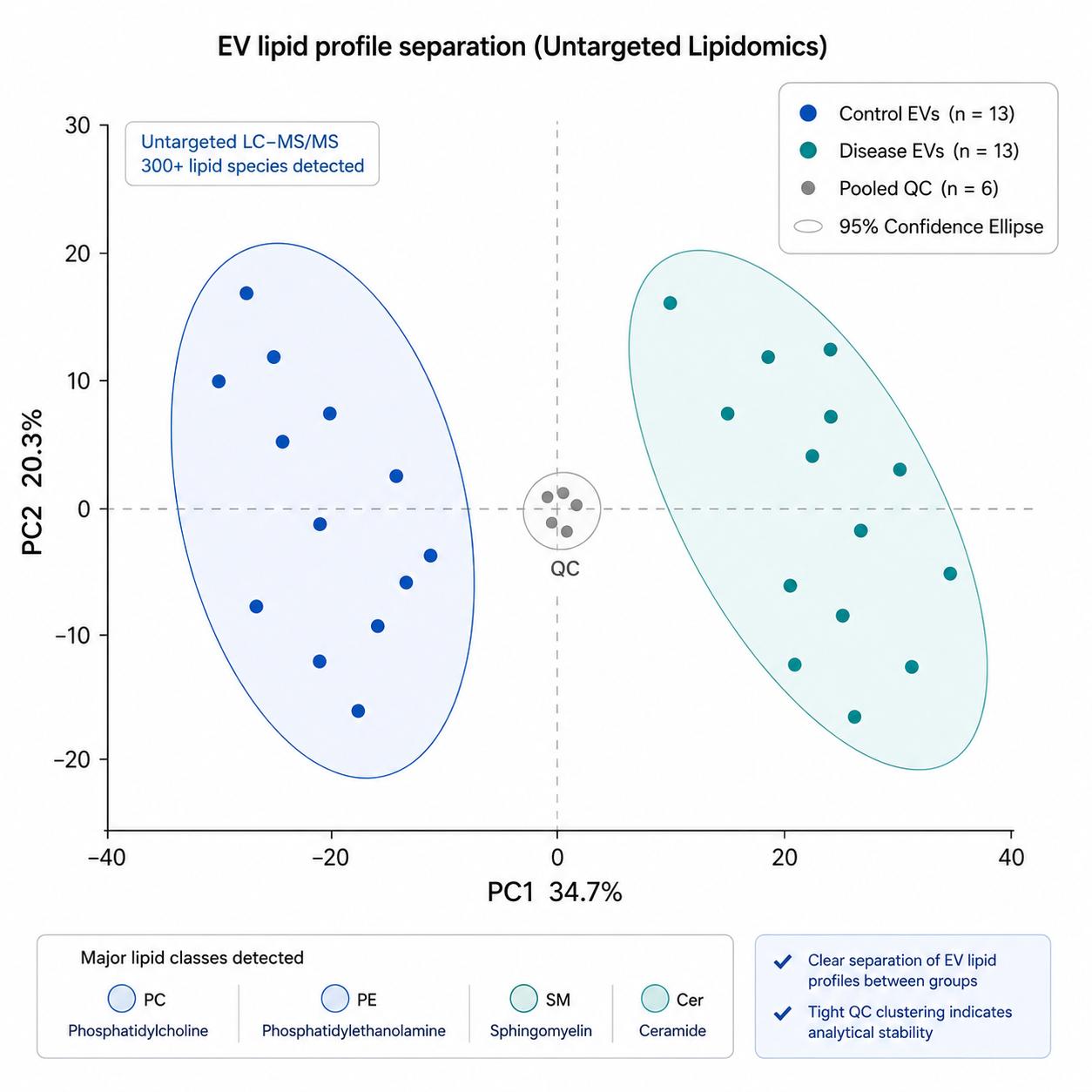
- PCA and PLS-DA with group separation statistics
QC metrics included:
- Pooled QC CV distribution, PCA with QC clustering, IS recovery
- EV characterization data: NTA, TEM, Western blot
Lipid class distribution in purified exosomes —
glycerophospholipids and sphingolipids dominate the EV lipidome.
PCA score plot of untargeted exosome lipidomics data showing
clear separation between experimental groups based on EV lipid profiles.
Targeted Quantification Report
Results provided:
- Absolute concentrations (nmol/mL or pmol/μg protein) with stable isotope IS
- 7-point calibration curves, r2, and back-calculated accuracy per batch
- Inter-batch CV and IS recovery tracking across the analytical sequence
EV Characterization & Pathway Analysis
- NTA, TEM, and Western blot data included with every project
- KEGG/Reactome pathway mapping with differential lipid overlay
- Publication-ready figures (TIFF/PDF, 600 dpi)
Explore our Lipidomics Solutions brochure to learn more about exosome lipidomics and our
complete extracellular vesicle analysis capabilities.
Download Brochure
Applications of Exosome Lipidomics
Tumor-Derived EV Biomarkers
Tumor EVs are enriched in phosphatidylserine and carry altered sphingomyelin-to-ceramide ratios
relative to healthy controls. Untargeted profiling identifies candidate lipid signatures without prior
hypothesis; targeted MRM validates them across independent cohorts.
CNS-Derived EVs in Neurological Disease
Neuron- and glia-derived EVs cross the blood-brain barrier into peripheral circulation. Ganglioside
profiles in CSF-derived EVs distinguish Alzheimer's and Parkinson's from controls; sulfatide
alterations in plasma EVs track with multiple sclerosis disease activity.
Metabolic Syndrome & Cardiovascular Risk
Adipocyte-derived EVs from insulin-resistant individuals carry elevated ceramide levels that correlate
with hepatic steatosis grade. Platelet-derived EVs in hyperlipidemia are enriched in arachidonic
acid-containing phospholipids, linking EV lipid cargo to thromboinflammatory risk. Endothelial EVs carry
distinct phospholipid signatures in early atherosclerosis that precede changes in plasma lipid profiles.
Drug Delivery Vehicle Engineering
The lipid composition of a drug carrier determines its cellular uptake, endosomal escape, and cargo
release — yet most LNPs are designed empirically. Comparative lipidomics of native EVs, engineered
liposomes, and clinical-stage LNPs identifies the compositional features that correlate with delivery
efficiency.
Sample Requirements for Exosome Lipidomics
| Sample Type |
Minimum Input |
Notes |
| Purified Exosomes / EVs |
10–50 μg protein |
Submit in PBS or Tris-buffered saline, pH 7.4. Provide isolation method details. Avoid detergents
(Triton X-100, SDS). Store at -80°C. |
| Plasma / Serum |
1–5 mL |
EDTA plasma preferred. Process within 2 h at 4°C. Pre-clear platelets (2,500×g) and microvesicles
(10,000×g). No heparin. Store at -80°C. |
| Cell Culture Supernatant |
10–50 mL |
EV-depleted FBS required (ultracentrifuged 100,000×g × 18 h). Record cell count, viability, and passage
number. Pre-clear by sequential centrifugation. |
| CSF |
1–2 mL |
Polypropylene tubes. Centrifuge 2,000×g × 10 min at 4°C. CSF EV concentration is low — larger volumes
improve lipid identification. |
| Urine |
10–50 mL |
Midstream collection with protease inhibitors. Centrifuge 2,000×g × 20 min. Tamm-Horsfall protein
polymers can trap EVs — brief DTT treatment before isolation may improve yield. |
FAQ — Exosome Lipidomics
Which approach should I use — untargeted or targeted?
Untargeted lipidomics is best for discovery — when you do not know which lipid classes or species are
altered in your system. It profiles 300+ species and identifies candidates for follow-up. Targeted MRM is
appropriate for validation — when you have a defined set of lipid species to quantify with absolute
concentrations across larger cohorts. Many EV projects use untargeted profiling in the discovery phase
(10–30 samples) followed by targeted validation of candidate markers in an independent cohort (50–200
samples). We offer both approaches and can transition discovery hits to targeted assays using the same
sample preparation workflow.
Which EV isolation method gives the cleanest lipidomics data?
SEC or UC with iodixanol density gradient polishing produce the cleanest EV preparations for lipidomics.
PEG-based precipitation kits co-isolate non-EV lipids and are not recommended for untargeted analysis. We
process samples from any isolation method — method-specific background subtraction and fraction-level
comparison are included.
Do I need to characterize my EVs before submitting?
EV characterization is included in our service. We perform NTA, TEM, and Western blot
(CD9/CD63/CD81/TSG101/calnexin) on every sample before lipid extraction. These data are included in the
final report — you will know the size, concentration, morphology, and purity of the EV population you
profiled.
Can you separate genuine EV lipids from lipoprotein contamination?
Yes — for SEC-isolated EVs, we profile early (EV-rich) and late (soluble/lipoprotein) fractions separately
and report lipids enriched in EV fractions. For UC samples, iodixanol gradient profiling separates EVs
(density ~1.10–1.18 g/mL) from lipoproteins. This fraction-level comparison is standard in every project.
How many replicates are recommended for EV lipidomics studies?
Minimum 5 biological replicates per group for in vivo studies, and 3 independent EV isolations for cell
culture experiments. Include matched control samples processed identically. Technical replicates (2–3
injections from the same extract) are run automatically as part of QC.
Is this service for clinical diagnostic use?
No. Our exosome lipidomics service is for research use only (RUO). It supports preclinical and
translational research — biomarker discovery, mechanism studies, and drug development — not clinical
diagnostics.